
A retinal cyst is a fluid-filled cavity inside the neurosensory retina, and the term covers several unrelated situations.
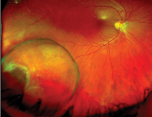
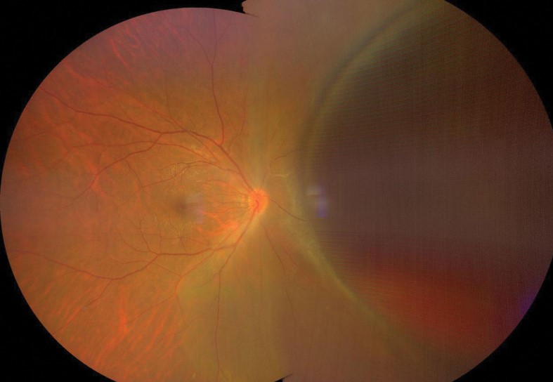
The one you meet most often is a peripheral, age-related change that nobody needs to treat.
The one you cannot afford to miss is the cystic space in the macula that signals active edema.
Peripheral microcystoid degeneration
Almost every adult has some of it.
Tiny, closely packed cysts form in the outer plexiform layer just behind the ora serrata, usually temporally, and they look like a fine, frosted or bubbly gray zone with sharp posterior borders on indirect ophthalmoscopy.
The old name, Blessig-Iwanoff cysts, still turns up in textbooks.
It is bilateral, symmetrical, and harmless on its own.
Its real importance is that it is the soil in which acquired retinoschisis grows.
Cysts coalesce, the inner wall thins, and a smooth dome appears, usually in the inferotemporal quadrant (see senile retinoschisis).
Cystic retinal tufts
These are small, white, elevated, round or oval excrescences of retinal tissue at the vitreous base.
They carry a small but real risk of a flap tear when the vitreous separates, which is why they are discussed alongside lattice degeneration (see retinal tuft and detachment risk).
Fundus Explorer Pro
Photograph the retinal findings described here with the phone already in your pocket — 22 D optics and built-in illumination in one handheld unit.
From Choroida — the team behind this siteMacular and posterior pole cysts
Cystic spaces in the macula are a different matter.
On OCT they appear as round, dark cavities in the inner nuclear and outer plexiform layers, and they mean that fluid is accumulating faster than the retina can clear it.
The usual causes are diabetic macular edema, retinal vein occlusion, uveitis, post-cataract surgery inflammation, retinitis pigmentosa, and tractional or epiretinal membrane disease (see cystoid macular edema).
Not every macular cyst is leakage.
In X-linked retinoschisis, in some optic pit maculopathies, and in several inherited conditions, the cavities are structural, and fluorescein angiography shows no petalloid leakage.
That single distinction, leakage versus no leakage, saves many patients from unnecessary injections.
Other types worth knowing
- Retinal pigment epithelial cysts, rare and usually stable
- Parasitic cysts, most notably cysticercosis, where a living scolex may be visible inside a translucent subretinal or vitreous cyst
- Congenital cysts in the setting of retinal dysplasia
- Macular microcysts after optic neuropathy, a finding that surprises people because it sits on the inner nuclear layer rather than the retinal vessels
How to examine and what to do
- Use scleral depression on indirect ophthalmoscopy for the periphery, and OCT for anything central
- Asymptomatic peripheral cysts need no treatment and no special follow-up beyond routine care
- A new cluster with symptoms of flashes or shadows should be checked for a tear
- Macular cysts need a diagnosis of the cause first, then treatment of that cause
What OCT added to the picture
Before OCT, the macular cyst was diagnosed from the stellate or honeycomb pattern at the fovea and from fluorescein leakage.
OCT showed that the cystic spaces sit at different depths depending on the cause.
In vascular edema, large cysts occupy the inner nuclear and outer plexiform layers and are often joined by a layer of subretinal fluid.
In traction, the cysts are smaller and the foveal contour is distorted.
In retinitis pigmentosa, cysts are common and may respond to topical carbonic anhydrase inhibitors, which is a different treatment pathway from the anti-VEGF route used in diabetic disease.
That difference in management is the reason to look carefully at the pattern, and not just report “cystoid macular edema” on the chart.
A simple approach in clinic
- Peripheral cysts found on routine dilated examination: reassure, note them, and move on
- Peripheral cysts with a convincing dome of retinoschisis: check for an outer-layer break and refer if one is present, since a retinoschisis detachment is rare but treatable
- Central cysts: obtain OCT, ask about recent eye surgery, uveitis, diabetes and drug exposure (niacin and taxanes cause non-leaking cystic maculopathy), and decide if angiography will add information
- Cysts in a child or adolescent, with no leakage on angiography: consider X-linked retinoschisis and refer for genetic confirmation
Counselling the patient
Patients who hear that they have "cysts in the retina" often imagine a tumor. It helps to explain that peripheral microcystoid change is a common ageing finding, that it does not spread, and that the only symptoms that matter are new flashes, a shower of floaters, or a shadow in the vision. Give those warning signs in writing, and advise that any such change needs a dilated examination within days, not weeks.
Prognosis
Peripheral cysts are benign.
Macular cysts follow the disease that produces them, and visual outcome depends on how long the retina has been swollen and whether the photoreceptor layer below stays intact.
Cysts that have been present for years leave Müller cell damage and a thin, disorganized inner retina, which is why early treatment of the cause produces better visual recovery than waiting to see whether the swelling resolves.


Document what you see
Two smartphone imaging tools built for everyday clinic use — one for the slit lamp, one for the fundus.
From Choroida — the team behind this siteReferences
- Byer NE. Clinical study of senile retinoschisis. Arch Ophthalmol. 1968;79:36-44.
- Shields JA, Shields CL. Intraocular Tumors: An Atlas and Textbook. 3rd ed. Philadelphia: Lippincott Williams & Wilkins; 2015.
- Ryan SJ, ed. Retina. 6th ed. London: Elsevier; 2018.
- Spaide RF. Retinal vascular cystoid macular edema: review and new theory. Retina. 2016;36:1823-1842.
