
A young woman with daily headaches and a normal brain scan can still be days away from permanent blindness.
Idiopathic intracranial hypertension (IIH) is raised intracranial pressure with no tumour, no hydrocephalus and no venous thrombosis to explain it.
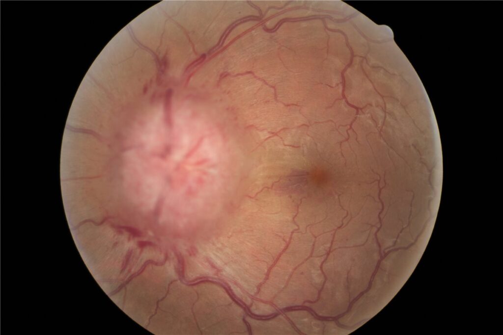
The pressure is transmitted along the optic nerve sheaths, and the optic discs swell.
Headache is what brings most patients in, but headache is not what threatens them.
Vision loss in IIH begins in the periphery and progresses silently, so a patient can lose a great deal of visual field while still reading the bottom line of the chart.
Recognising IIH, grading the papilledema, and monitoring visual fields rather than acuity is what actually protects sight in this disease.
What Is Idiopathic Intracranial Hypertension?
Idiopathic intracranial hypertension (IIH) is a syndrome of raised cerebrospinal fluid pressure occurring without an identifiable structural, vascular or infectious cause.
Diagnosis rests on the modified Dandy criteria:
- Signs and symptoms of raised intracranial pressure, most often headache and papilledema
- No localising neurological signs, with the recognised exception of a sixth nerve palsy
- Normal neuroimaging, including venography to exclude cerebral venous sinus thrombosis
- Raised opening pressure on lumbar puncture with normal cerebrospinal fluid composition
- No other identified cause of raised intracranial pressure
The word “idiopathic” carries real weight here: IIH is a diagnosis of exclusion, and the imaging done to reach it is as much a part of the diagnosis as the lumbar puncture.
Epidemiology
IIH has a strikingly consistent demographic pattern that is itself a diagnostic clue.
- It occurs predominantly in women of childbearing age
- Obesity, and particularly recent weight gain, is the strongest and most consistently reported association
- Incidence tracks population obesity rates, so the burden of the disease has been rising
- It can occur in men, in children and in patients who are not obese, and these atypical presentations deserve a lower threshold for hunting a secondary cause
An older patient, a man, or a thin patient with apparent IIH should prompt a harder look for venous sinus thrombosis, medication effect or another secondary cause before the label is accepted.
Fundus Explorer Pro
Photograph the retinal findings described here with the phone already in your pocket — 22 D optics and built-in illumination in one handheld unit.
From Choroida — the team behind this sitePathophysiology
The precise mechanism remains incompletely understood, but the sequence that damages vision is well characterised.
- Cerebrospinal fluid pressure rises, whether through impaired CSF absorption, altered venous outflow or as yet undefined metabolic factors
- Raised pressure is transmitted forward along the subarachnoid space surrounding each optic nerve
- Axoplasmic flow within retinal ganglion cell axons stalls at the lamina cribrosa, and the accumulated axoplasm produces the visible disc swelling
- Persistent swelling compresses axons and compromises the capillary supply at the disc
- Chronic, untreated papilledema culminates in axonal loss and secondary optic atrophy, at which point vision loss is permanent
This is why disc swelling that finally “settles” in an untreated patient is an ominous sign rather than a reassuring one: the swelling can resolve simply because there are no longer enough axons left to swell.
Risk Factors
Patient-Related Risk Factors
- Female sex and childbearing age
- Obesity, and especially a recent significant increase in weight
- Polycystic ovary syndrome and other endocrine disturbances
Medication and Secondary Associations
- Tetracycline-class antibiotics, including doxycycline and minocycline
- Vitamin A derivatives such as isotretinoin, and excessive vitamin A intake
- Corticosteroid withdrawal
- Cerebral venous sinus thrombosis and other venous outflow obstruction, which produce a clinically identical picture but are by definition not idiopathic
A careful medication history is one of the highest-yield parts of the consultation, because a drug-induced case can resolve simply by stopping the offending agent.
Clinical Presentation
Symptoms
- Headache, typically daily, often worse on waking or on lying flat
- Transient visual obscurations – brief greyouts of vision lasting seconds, often provoked by standing or bending
- Pulsatile tinnitus, described as a whooshing sound in time with the pulse
- Horizontal double vision from a sixth nerve palsy, a false localising sign of raised pressure
- Progressive peripheral field loss, which patients frequently do not notice until it is advanced
Examination Findings
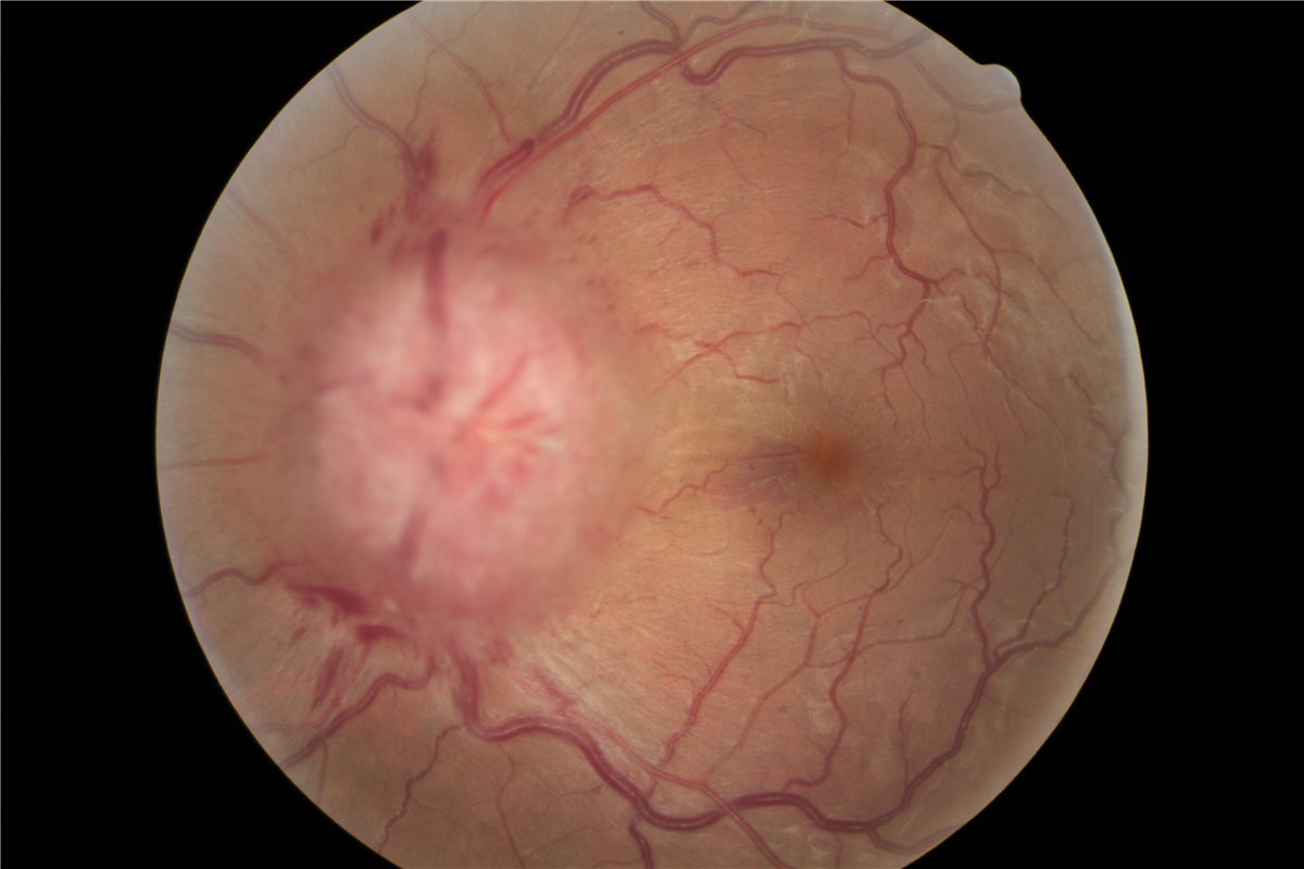
- Bilateral papilledema – an elevated optic disc with blurred margins and obscuration of vessels as they cross the disc edge
- Peripapillary haemorrhages and cotton-wool spots in more severe swelling
- Enlargement of the physiological blind spot, often the earliest measurable field defect
- Peripheral and nasal visual field constriction as the disease progresses
- Preserved central visual acuity until late, which is precisely why acuity is a poor monitoring tool
- Possible sixth nerve palsy with limited abduction
Papilledema in IIH is almost always bilateral, so a genuinely unilateral swollen disc should redirect the workup toward a different diagnosis entirely.
Diagnostic Evaluation
Neuroimaging
- MRI of the brain to exclude a mass lesion, hydrocephalus or meningeal disease
- MR venography to exclude cerebral venous sinus thrombosis, which is mandatory rather than optional
- Supportive imaging signs of raised pressure include an empty sella, flattening of the posterior globe, distended optic nerve sheaths and transverse sinus stenosis
Lumbar Puncture
- Performed only after imaging has excluded a mass lesion
- Documents a raised opening pressure, measured in the lateral decubitus position with the legs extended
- Cerebrospinal fluid composition must be normal – abnormal constituents point to an inflammatory or infectious cause instead
Ophthalmic Monitoring
- Formal automated perimetry is the primary tool for detecting and tracking visual loss
- Disc photography and OCT of the retinal nerve fibre layer to grade and follow the degree of swelling objectively
- Serial assessment at intervals dictated by severity, since deterioration can be rapid in fulminant disease
Visual fields, not visual acuity, drive management decisions in IIH – a patient with 6/6 vision and progressive field constriction is losing the disease.
Differential Diagnosis
Conditions that can produce a similar picture include:
- Cerebral venous sinus thrombosis – identical clinical syndrome, excluded only by venography
- Intracranial mass lesion or hydrocephalus – excluded on MRI
- Medication-induced intracranial hypertension – the same syndrome with an identifiable drug trigger
- Malignant hypertension – severe systemic hypertension producing disc swelling and retinopathy
- Pseudopapilledema from optic disc drusen – elevated discs without true swelling, distinguished on ultrasound, autofluorescence or OCT
- Bilateral optic neuritis or infiltrative optic neuropathy – usually with early, marked loss of acuity and colour vision
Separating true papilledema from pseudopapilledema early avoids both an unnecessary lumbar puncture and, in the opposite error, a dangerous false reassurance.
Management
Weight Management
- Sustained weight loss is disease-modifying rather than merely supportive, and can produce remission
- Structured weight management support should be offered early rather than deferred
- Bariatric surgery is an option in appropriately selected patients with refractory disease
Medical Therapy
- Acetazolamide is first-line therapy; the Idiopathic Intracranial Hypertension Treatment Trial showed improved visual field function when it was combined with a weight-reduction diet in mild visual loss
- Topiramate is an alternative, with the practical advantage of promoting weight loss alongside its effect on pressure
- Headache management in parallel, since headache often persists even after papilledema resolves
- Withdrawal of any implicated medication
Surgical and Interventional Options
- Optic nerve sheath fenestration, directed primarily at protecting vision when visual loss is the dominant problem
- Cerebrospinal fluid diversion by ventriculoperitoneal or lumboperitoneal shunting, particularly when headache and pressure symptoms dominate
- Venous sinus stenting in selected patients with significant transverse sinus stenosis and a measured pressure gradient
- Urgent intervention for fulminant IIH, where severe visual loss develops over days
The choice between these procedures turns on whether vision or headache is the primary threat, and fulminant disease is a surgical emergency rather than a medication decision.
Prognosis
Outcome depends heavily on how early treatment starts and how closely vision is monitored.
- Most patients treated promptly retain good vision, and papilledema resolves as pressure is controlled
- Sustained weight loss is associated with durable remission in many patients
- Recurrence is well recognised, particularly with weight regain, so discharge should not be automatic once the discs flatten
- Untreated or fulminant disease can cause irreversible optic atrophy and severe permanent visual loss
- Headache frequently outlasts the papilledema and often needs its own long-term management
The distinguishing feature of IIH is that a fully preventable blindness sits alongside a symptom – headache – that dominates the consultation but is not the thing most likely to harm the patient.


Document what you see
Two smartphone imaging tools built for everyday clinic use — one for the slit lamp, one for the fundus.
From Choroida — the team behind this siteReferences
- Friedman DI, Liu GT, Digre KB. Revised diagnostic criteria for the pseudotumor cerebri syndrome in adults and children. Neurology. 2013.
- NORDIC Idiopathic Intracranial Hypertension Study Group. Effect of acetazolamide on visual function in patients with idiopathic intracranial hypertension and mild visual loss: the IIH Treatment Trial. JAMA. 2014.
- Mollan SP, Davies B, Silver NC, et al. Idiopathic intracranial hypertension: consensus guidelines on management. Journal of Neurology, Neurosurgery & Psychiatry. 2018.
- Markey KA, Mollan SP, Jensen RH, Sinclair AJ. Understanding idiopathic intracranial hypertension: mechanisms, management, and future directions. The Lancet Neurology. 2016.
- Idiopathic Intracranial Hypertension. EyeWiki, American Academy of Ophthalmology.
Test yourself
A few questions straight from this article.
-
Which focal neurological sign is still compatible with idiopathic intracranial hypertension under the modified Dandy criteria?
The modified Dandy criteria require no localising neurological signs, with the recognised exception of a sixth nerve palsy, which is a false localising sign of raised pressure. -
Which imaging study is mandatory before accepting a diagnosis of idiopathic intracranial hypertension?
Cerebral venous sinus thrombosis produces an identical clinical syndrome and is excluded only by venography, so MR venography is mandatory rather than optional. -
What mechanism produces the visible optic disc swelling in idiopathic intracranial hypertension?
Pressure transmitted along the optic nerve subarachnoid space stalls axoplasmic flow within ganglion cell axons at the lamina cribrosa, and the accumulated axoplasm produces the swelling. -
Why is spontaneously settling papilledema an ominous sign in an untreated IIH patient?
Chronic untreated papilledema ends in axonal loss and secondary optic atrophy, so swelling can resolve simply because there are no longer enough axons left to swell. -
Which factor is the strongest and most consistently reported association with idiopathic intracranial hypertension?
IIH occurs predominantly in women of childbearing age, and obesity, especially a recent significant increase in weight, is its strongest and most consistently reported association. -
Which medication group is a recognised trigger for intracranial hypertension?
Tetracyclines including doxycycline and minocycline, vitamin A derivatives such as isotretinoin, and corticosteroid withdrawal are recognised triggers, so a drug-induced case may resolve by stopping the agent. -
Which test should drive monitoring decisions in idiopathic intracranial hypertension?
Central acuity is preserved until late, so formal automated perimetry is the primary tool for detecting and tracking visual loss, and fields rather than acuity drive management. -
Which drug is first-line medical therapy for idiopathic intracranial hypertension?
Acetazolamide is first-line, and the Idiopathic Intracranial Hypertension Treatment Trial showed improved visual field function when it was combined with a weight-reduction diet in mild visual loss. -
Which procedure is directed primarily at protecting vision when visual loss dominates in IIH?
Optic nerve sheath fenestration is aimed primarily at protecting vision, while cerebrospinal fluid diversion by shunting is favoured when headache and pressure symptoms dominate. -
Which outcome is characteristic of idiopathic intracranial hypertension after papilledema resolves?
Headache frequently outlasts the papilledema and often needs long-term management of its own, and recurrence is well recognised with weight regain, so discharge should not be automatic.

Writes and reviews the clinical articles on Educate. Every post is checked against peer-reviewed sources before it goes live.


