
In a cornea with stromal keratitis, a ring of gray-white infiltrate may form around a central focus, with a clear zone in between.
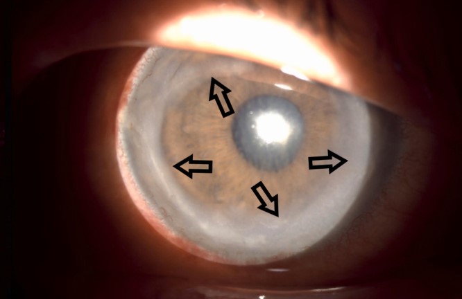
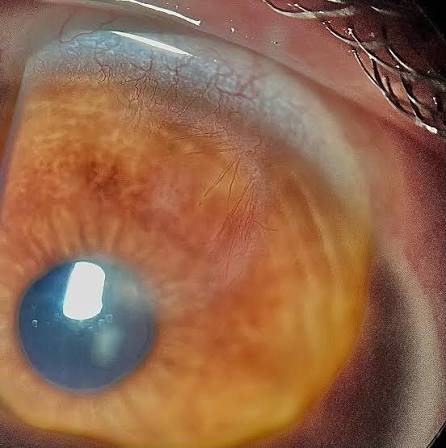
This is a Wessely immune ring, and it represents a localized immune reaction in the corneal stroma.
It is a clue to the nature of the infection, and it is a sign of significant antigenic stimulation.
How it forms
When antigen is released in the center of the stroma, as from a replicating virus or an organism, antibodies diffuse in from the limbal vessels.
The two meet at a zone of equivalence, where they precipitate and attract neutrophils and complement.
The result is an annular band of inflammation and stromal opacity, concentric with the original focus.
A clear zone between the center and the ring corresponds to the region of antigen excess.
The ring was described by Karl Wessely in 1911 in experimental work on rabbit corneas.
When you see it
- Herpes simplex stromal keratitis, in which it occurs in necrotizing disease
- Fungal keratitis, where it may appear around a satellite lesion
- Acanthamoeba keratitis, in which a ring-shaped infiltrate is a well-known late sign (see Acanthamoeba ring infiltrate)
- Bacterial keratitis, particularly Pseudomonas, with ring ulcer
- Reaction to foreign material, for example, after a retained foreign body or a corneal injection
- Vaccinia and some other viral infections
All-fit Slit-Lamp Adapter
Record and share exactly what you see at the slit lamp. One adapter fits any slit lamp or surgical microscope — and any smartphone.
From Choroida — the team behind this siteHow it differs from other rings
A ring ulcer of staphylococcal or marginal keratitis lies near the limbus.
The Wessely ring lies in the stroma, within the central or paracentral cornea, with a clear zone between the center and the ring.
A Fleischer ring is an iron line around a keratoconus cone, and a Kayser-Fleischer ring is copper deposition at the periphery in Wilson disease.
The Coats white ring is a small, white, iron-containing ring that forms around an old foreign body.
What it means clinically
A ring signals active, intense stromal inflammation, and it often means a more severe or longer-standing disease.
In Acanthamoeba keratitis it may suggest delay in diagnosis.
In herpetic stromal keratitis, it is a sign of necrotizing disease that needs immune suppression along with antiviral cover (see herpetic stromal keratitis).
Evaluation
- Detailed history of contact lens wear, trauma, herpes, and previous treatment
- Slit-lamp assessment of the ring, central focus, epithelial defect, and endothelial involvement
- Corneal scraping for smears, cultures, and PCR, including for Acanthamoeba when relevant (see corneal ulcer culture technique)
- Confocal microscopy, when available, to look for cysts or hyphae
Treatment
Treatment follows the cause.
Antiviral therapy and corticosteroids are used for herpetic disease, with antimicrobial therapy for bacterial or fungal infection.
Acanthamoeba requires prolonged therapy with biguanides and diamidines.
Steroids must be used with great caution unless the cause is clear.
Recognizing the ring at the slit lamp
The ring is a gray-white, circular band in the stroma, usually 2 to 4 mm in diameter, concentric with a central focus of infection or foreign material. Between the center and the ring there is a zone of relatively clear cornea. The overlying epithelium may be intact or ulcerated, and the anterior chamber often shows cells, flare, and sometimes a hypopyon. Look for stromal edema, new vessels, and thinning, which signal severe disease.
What the ring suggests about the cause
In herpetic stromal keratitis, a ring can accompany necrotizing disease and often follows repeated attacks. In Acanthamoeba keratitis, the ring appears later in the course, and the disease is typically very painful. In fungal keratitis, rings are associated with deep stromal infiltrates and satellite lesions. A ring around a foreign body suggests a reaction to material in the stroma. No single feature is diagnostic, so scraping and culture, PCR, and sometimes biopsy are needed.
Practical management points
Take cultures before treatment where possible, and avoid steroids until infection has been excluded or controlled. Monitor closely for thinning and perforation, and be prepared to arrange transplantation if the cornea melts. Educate the patient about the importance of regular drops, follow-up, and avoiding contact lens use until the eye has healed.
Pitfalls
- Do not assume that every ring is Acanthamoeba, because herpes and bacteria can produce similar rings
- Do not start steroids before cultures when infection is possible
- Do not forget to ask about contact lens history, water exposure, and previous herpes
- Do not rely on a single negative scraping in a typical case, since repeat samples or confocal microscopy may reveal the organism
Prognosis
A Wessely ring often leaves a stromal scar.
Visual outcome depends on the cause, the speed of diagnosis, and the response to treatment.


Document what you see
Two smartphone imaging tools built for everyday clinic use — one for the slit lamp, one for the fundus.
From Choroida — the team behind this siteReferences
- Wessely K. Ueber anaphylaktische Erscheinungen an der Hornhaut. Muench Med Wochenschr. 1911;58:1713-1714.
- Krachmer JH, Mannis MJ, Holland EJ, eds. Cornea. 3rd ed. Philadelphia: Elsevier; 2011.
- Lorenzo-Morales J, Khan NA, Walochnik J. An update on Acanthamoeba keratitis: diagnosis, pathogenesis and treatment. Parasite. 2015;22:10.
- Holland EJ, Schwartz GS. Classification of herpes simplex virus keratitis. Cornea. 1999;18:144-154.
