
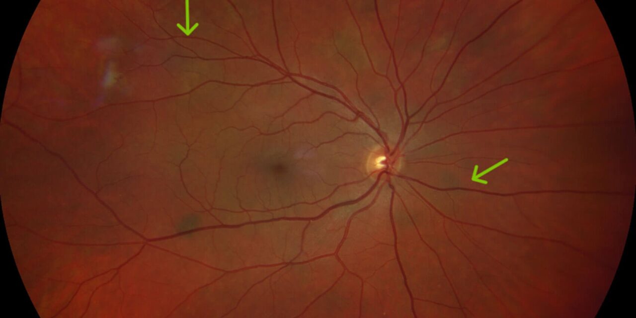
Neurofibromatosis type 2 is genetically and clinically distinct from the more commonly encountered neurofibromatosis type 1, and its ocular manifestations reflect this distinction, centering on an early, distinctive form of cataract and specific retinal findings rather than the iris Lisch nodules that are the hallmark ocular finding of type 1.
Genetics and Systemic Features
Neurofibromatosis type 2 results from mutations in the NF2 gene, which encodes a protein called merlin, and is characterized primarily by bilateral vestibular schwannomas, benign tumors of the eighth cranial nerve that typically cause progressive hearing loss and can affect balance, along with a tendency toward other central and peripheral nervous system tumors, including meningiomas and spinal schwannomas.
Ocular Findings
Cataract
A distinctive, early-onset cataract, often of a posterior subcapsular or cortical type, is one of the most characteristic and clinically useful ocular findings in neurofibromatosis type 2, and its early onset, sometimes in childhood or young adulthood, well before the age typical of age-related cataract, can be a valuable diagnostic clue, occasionally preceding the diagnosis of the systemic syndrome itself.
Epiretinal Membrane
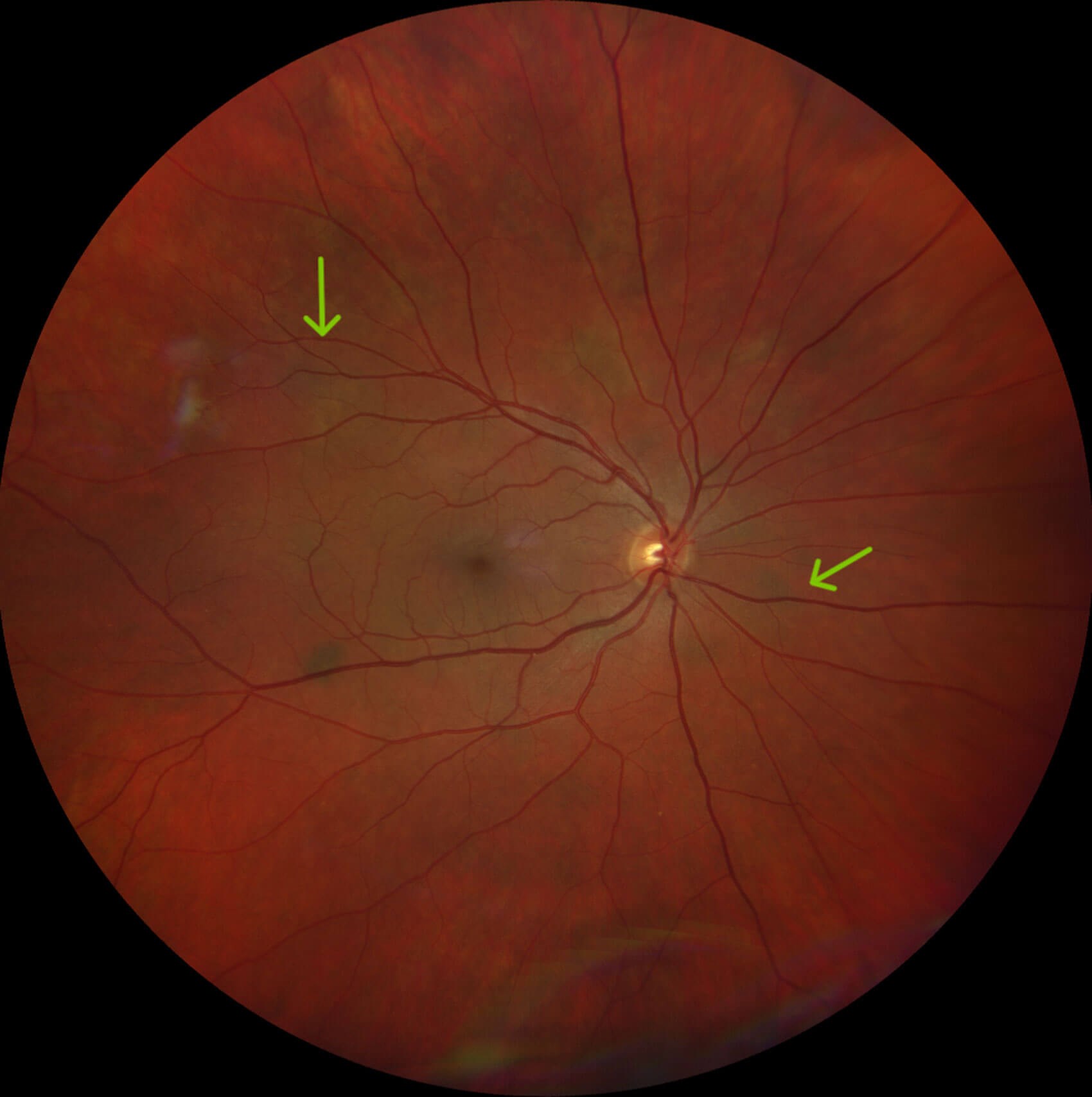
Epiretinal membranes, thin fibrous membranes that can form on the retinal surface and cause visual distortion or blurring, occur with increased frequency in neurofibromatosis type 2 and can, in some patients, warrant surgical removal if visually significant.
Combined Hamartoma of the Retina and Retinal Pigment Epithelium
This distinctive lesion, involving a combined proliferation of retinal and retinal pigment epithelial tissue, has been associated with neurofibromatosis type 2, and it can cause visual distortion or reduced vision depending on its location, particularly if it involves the macula.
Optic Nerve Sheath Meningioma
Given the broader tendency toward meningioma formation in this syndrome, optic nerve sheath meningiomas can occur, potentially threatening vision through direct optic nerve compression, and require specific neuro-ophthalmic evaluation and monitoring when suspected.
Fundus Explorer Pro
Photograph the retinal findings described here with the phone already in your pocket — 22 D optics and built-in illumination in one handheld unit.
From Choroida — the team behind this siteDistinguishing From Neurofibromatosis Type 1
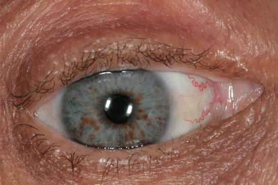
- Lisch nodules, the hallmark iris finding of neurofibromatosis type 1, are not a typical feature of type 2, and their absence should not be reassuring against an NF2 diagnosis when other suggestive features, particularly early cataract, are present
- Optic pathway glioma, common in type 1, is not a typical feature of type 2
- The characteristic early cataract and epiretinal changes of type 2 are, conversely, not typical features of type 1
Recognizing these distinctions is clinically important, since the two conditions carry different systemic surveillance needs, genetic counseling implications, and inheritance considerations, despite the similarity in name.
Diagnostic and Screening Role of Ophthalmic Findings
Because the characteristic early cataract of neurofibromatosis type 2 can appear before other systemic manifestations, particularly in children or young adults from a family with a known history of the condition, ophthalmic examination can play a genuine role in early recognition, prompting further evaluation, including audiologic assessment and neuroimaging for vestibular schwannoma, in an at-risk individual.
Management
Cataract Management
Cataract surgery is performed using standard techniques when visually significant, with attention to the sometimes atypical lens changes and any associated ocular findings that could affect surgical planning.
Monitoring for Other Ocular Findings
Regular ophthalmic surveillance, including dilated fundus examination for epiretinal membrane, combined hamartoma, and other retinal findings, along with neuro-ophthalmic assessment when optic nerve involvement is a concern, is part of the comprehensive, coordinated care these patients need.
Coordinated Systemic Care
Given the significant systemic implications of neurofibromatosis type 2, including progressive hearing loss and the risk of multiple central nervous system tumors, ophthalmic care is best integrated with neurology, otolaryngology, and neurosurgery as part of a coordinated, multidisciplinary management plan.
Prognosis
Ocular findings in neurofibromatosis type 2 are generally manageable, with cataract surgery and, when needed, epiretinal membrane surgery providing good visual outcomes, though ongoing surveillance for new or progressive ocular findings remains important given the chronic, evolving nature of this syndrome.


Document what you see
Two smartphone imaging tools built for everyday clinic use — one for the slit lamp, one for the fundus.
From Choroida — the team behind this siteReferences
- Ragge NK, Baser ME, Klein J, et al. Ocular abnormalities in neurofibromatosis 2. Am J Ophthalmol. 1995;120:634-641.
- Evans DG, Huson SM, Donnai D, et al. A clinical study of type 2 neurofibromatosis. Q J Med. 1992;84:603-618.
- Landau K, Yasargil GM. Ocular fundus in neurofibromatosis type 2. Br J Ophthalmol. 1995;79:1015-1017.
- Bosch MM, Boltshauser E, Harpes P, Landau K. Ophthalmologic findings and long-term course in patients with neurofibromatosis type 2. Am J Ophthalmol. 2006;141:1068-1077.
