
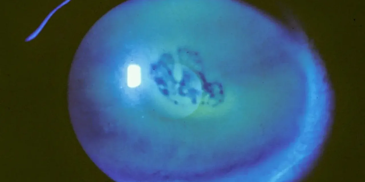
Recurrent corneal erosion syndrome is characterized by episodes of spontaneous corneal epithelial breakdown, classically occurring on waking, caused by poor adhesion between the epithelium and its underlying basement membrane at a site of prior injury or underlying dystrophy.
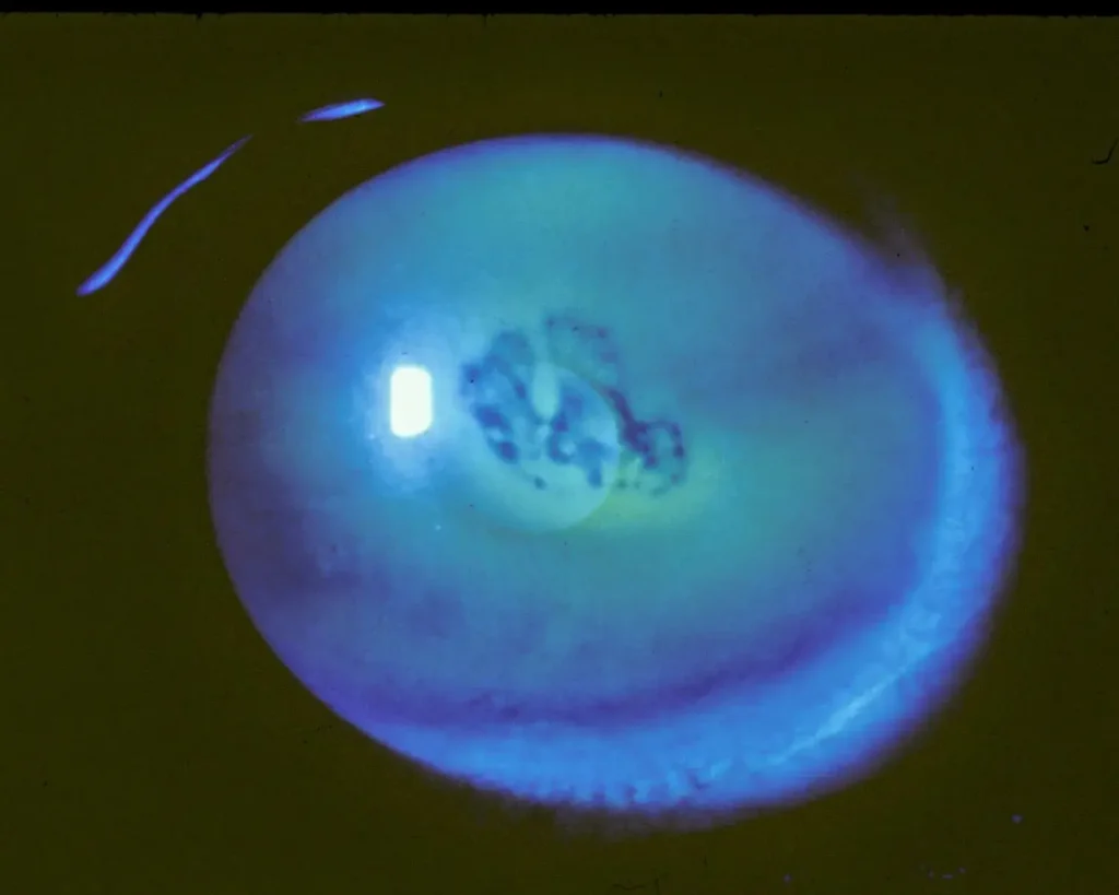
The defining feature is repetition — a single corneal abrasion is common and unremarkable, but the same small area breaking down again and again, often months or years after the original injury has apparently healed, is what defines this syndrome and points to a persistent structural problem rather than a series of unrelated new injuries.
Why Mornings
During sleep, the eyelids provide less oxygen and moisture exchange than during waking hours, and the epithelium can become mildly edematous and adherent to the undersurface of the eyelid overnight.
When the eyes open and the lids move across the ocular surface with the first blinks of the morning, a poorly adherent area of epithelium — one that never properly re-established firm attachment to its basement membrane after a prior injury — can be mechanically sheared away.
This is why the hallmark presentation is a sudden, sharp pain on waking or shortly after, often severe enough that patients recall the exact moment it happened.
Underlying Causes
- Prior traumatic corneal abrasion — particularly from a fingernail, paper edge, tree branch, or other sharp organic material, injuries that seem to disproportionately predispose to recurrent erosion compared with other mechanisms
- Epithelial basement membrane dystrophy (map-dot-fingerprint dystrophy) — an inherited or acquired abnormality of the basement membrane itself, present in a substantial proportion of patients with recurrent erosion even without a clear preceding traumatic injury
- Other anterior corneal dystrophies affecting epithelial adhesion
- Prior corneal surgery, including photorefractive keratectomy or other procedures involving the epithelium
All-fit Slit-Lamp Adapter
Record and share exactly what you see at the slit lamp. One adapter fits any slit lamp or surgical microscope — and any smartphone.
From Choroida — the team behind this siteClinical Presentation
Patients describe sudden, sharp eye pain typically occurring on waking or shortly after, accompanied by tearing, photophobia, and blurred vision, sometimes severe enough to prompt an urgent visit.
Between episodes, many patients are entirely asymptomatic, and the eye can appear completely normal on exam if seen when not actively erosing.
This is why a good history covering the pattern of recurrent episodes — not just the current presentation — is essential to reaching the diagnosis.
A patient who has been seen for “recurrent abrasions” by several different clinicians over time, each visit treated as an isolated new injury, has often not had the underlying recurrent pattern actually recognized and named, which is why explicitly asking about prior similar episodes at any visit for a corneal abrasion is genuinely worthwhile.
Exam Findings
- During an active episode: a frank epithelial defect, often at or near the site of a prior injury, with fluorescein staining
- Between episodes: subtle findings that are easy to miss without deliberate, careful examination — epithelial irregularity, microcysts, fine map-like lines, or negative staining patterns after fluorescein instillation and a brief wait, corresponding to areas of poor epithelial adherence
- Signs of epithelial basement membrane dystrophy if present — map lines, dot-like cysts, or fingerprint-like ridges, sometimes bilateral even when only one eye has been symptomatic
- A negative fluorescein staining “pooling” pattern that can help identify loose epithelium even without an active defect, useful for confirming the diagnosis and planning treatment between acute episodes
Differential Diagnosis
- Neurotrophic keratopathy — reduced corneal sensation and typically less acute pain relative to the degree of epithelial breakdown, a different underlying mechanism
- Dry eye disease with recurrent punctate keratopathy — more diffuse, chronic surface irregularity rather than a discrete, recurring defect at a consistent location
- Photokeratitis — bilateral, tied to a specific recent UV exposure history rather than a pattern of recurrence over months to years
- Herpetic keratitis — a dendritic pattern rather than the more geographic or irregular defect typical of erosion, and generally without the same clear history of an antecedent traumatic injury
Management
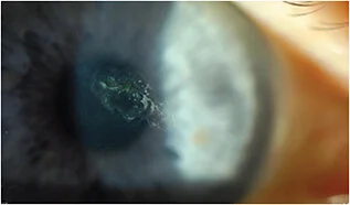
Acute episodes are treated similarly to any traumatic corneal abrasion: lubrication, cycloplegia for comfort, topical antibiotic prophylaxis, and a bandage contact lens in more severe or slow-healing cases, prioritizing comfort and infection prevention while the acute defect re-epithelializes.
The real focus of management, however, is preventing recurrence: aggressive lubrication with preservative-free artificial tears through the day and a thicker lubricating ointment at bedtime specifically to reduce the overnight adhesion problem is first-line, often continued for months even after symptoms have settled, because stopping too early commonly leads to relapse.
For patients who continue to have recurrent episodes despite consistent lubrication, hypertonic saline drops or ointment can help by reducing epithelial edema, and represents a reasonable intermediate step before considering a more invasive, definitive procedure.
More definitive interventions — anterior stromal micropuncture, diamond burr epithelial debridement, or phototherapeutic keratectomy — are used for refractory cases, working by promoting stronger, more durable adhesion complexes between the epithelium and basement membrane at the site of chronic poor attachment.
The choice among these procedures depends on the location, extent, and underlying cause of the erosion, with central, visually significant areas requiring more careful technique selection than peripheral ones.
Most patients who progress to these more definitive procedures achieve durable, lasting relief, which is worth emphasizing to a patient who may have already endured years of intermittent, disruptive episodes before reaching this stage of treatment.


Document what you see
Two smartphone imaging tools built for everyday clinic use — one for the slit lamp, one for the fundus.
From Choroida — the team behind this siteReferences
- Reidy JJ, Paulus MP, Gona S. Recurrent erosions of the cornea: epidemiology and treatment. Cornea.
- Ramamurthi S, Rahman MQ, Dutton GN, Ramaesh K. Pathogenesis, clinical features and management of recurrent corneal erosions. Eye.
- American Academy of Ophthalmology. Basic and Clinical Science Course, Section 8: External Disease and Cornea.
Test yourself
A few questions straight from this article.
-
What structural problem underlies recurrent corneal erosion syndrome?
The syndrome is defined by repeated spontaneous epithelial breakdown at a site where the epithelium never re-established firm attachment to its underlying basement membrane. -
Why does recurrent corneal erosion classically cause pain on waking?
During sleep the epithelium becomes mildly oedematous and adherent to the undersurface of the eyelid; the first lid movements of the morning mechanically strip the poorly attached area away. -
Which injury type disproportionately predisposes to recurrent corneal erosion?
The article singles out abrasions from a fingernail, paper edge or tree branch as seeming to predispose to recurrent erosion far more than other injury mechanisms. -
Which dystrophy commonly underlies recurrent corneal erosion without any preceding trauma?
Map-dot-fingerprint dystrophy is an abnormality of the basement membrane itself and is present in a substantial proportion of erosion patients who report no clear preceding injury. -
Which fluorescein finding helps identify loose epithelium in recurrent corneal erosion between attacks?
Negative staining marks areas of poor epithelial adherence and can confirm the diagnosis and guide treatment planning even when no active defect is present. -
Which feature distinguishes neurotrophic keratopathy from recurrent corneal erosion?
Neurotrophic keratopathy shows diminished corneal sensation and typically far less acute pain than the degree of epithelial breakdown would suggest — a different underlying mechanism entirely. -
How is an acute episode of recurrent corneal erosion treated?
The acute episode is managed exactly like any traumatic abrasion, prioritising comfort and infection prevention while the defect re-epithelialises; a bandage contact lens is added in severe or slow-healing cases. -
What is first-line treatment for preventing recurrence in recurrent corneal erosion?
Aggressive lubrication with a thicker ointment at bedtime specifically targets the overnight adhesion problem, and is often continued for months because stopping early commonly leads to relapse. -
Which treatment is a reasonable intermediate step before invasive procedures in recurrent corneal erosion?
Hypertonic saline reduces epithelial oedema and is offered to patients who keep eroding despite consistent lubrication, before moving to a more invasive definitive procedure. -
What outcome follows definitive procedures for refractory recurrent corneal erosion?
Micropuncture, diamond burr debridement and phototherapeutic keratectomy work by promoting stronger adhesion complexes, and most patients reaching this stage obtain lasting relief — worth telling someone who has endured years of episodes.
