
The cornea was the first human tissue ever transplanted successfully, and it remains the most transplanted tissue in the world.
Penetrating keratoplasty (PK) replaces the full thickness of the cornea with donor tissue.
For most of the last century it was the only corneal transplant on offer.
Lamellar techniques have since taken over much of its former workload, because replacing only the diseased layer is safer and heals faster.
But PK has not disappeared – it remains the operation when the full thickness of the cornea is damaged, and nothing else will do.
Knowing when full-thickness surgery is still the right answer, and how to recognise rejection early, is essential for anyone following these patients.
What Is Penetrating Keratoplasty?
Penetrating keratoplasty is full-thickness corneal transplantation, in which a circular disc of the patient’s cornea is removed and replaced with a matching donor button secured by sutures.
It sits within a family of corneal transplant procedures:
- Penetrating keratoplasty (PK) – all corneal layers replaced
- Deep anterior lamellar keratoplasty (DALK) – stroma replaced, the patient’s own endothelium retained
- Descemet stripping automated endothelial keratoplasty (DSAEK) – posterior stroma and endothelium replaced
- Descemet membrane endothelial keratoplasty (DMEK) – Descemet membrane and endothelium alone replaced
The general principle of modern corneal surgery is to replace only what is diseased, which is why PK is now reserved for full-thickness pathology.
Epidemiology
Corneal transplantation is by a wide margin the most commonly performed transplant procedure worldwide.
- The cornea is avascular and immune-privileged, which is why graft survival compares favourably with solid organ transplantation and systemic immunosuppression is not routinely required
- The proportion of transplants performed as PK has fallen substantially as endothelial and lamellar techniques have been adopted
- Endothelial failure – Fuchs dystrophy and pseudophakic bullous keratopathy – was historically the leading indication and is now largely managed by DSAEK or DMEK
- Keratoconus, once a common PK indication, is increasingly treated with DALK or stabilised earlier with corneal cross-linking
- Access to donor tissue remains the principal limiting factor in many parts of the world
The shift away from PK is one of the clearest examples in ophthalmology of a procedure being narrowed rather than abandoned as better-targeted alternatives arrived.
All-fit Slit-Lamp Adapter
Record and share exactly what you see at the slit lamp. One adapter fits any slit lamp or surgical microscope — and any smartphone.
From Choroida — the team behind this siteIndications
When Full-Thickness Replacement Is Needed
- Full-thickness corneal scarring, whether from infection, trauma or chemical injury
- Advanced keratoconus with significant scarring or previous hydrops, where DALK is not feasible
- Failed previous grafts
- Corneal perforation or descemetocele requiring tectonic support to restore globe integrity
- Severe infectious keratitis unresponsive to medical therapy – a therapeutic rather than optical graft
- Corneal dystrophies affecting all layers
When a Lamellar Procedure Is Preferred
- Isolated endothelial disease – Fuchs dystrophy, pseudophakic bullous keratopathy – where DSAEK or DMEK gives faster recovery and lower rejection risk
- Anterior stromal disease with a healthy endothelium, where DALK avoids the risk of endothelial rejection entirely
The decisive question at the planning stage is whether the patient’s own endothelium is healthy – if it is, a lamellar procedure is almost always the better operation.
Surgical Principles
The operation follows a well-established sequence.
- Donor tissue is assessed for endothelial cell density and clarity before use
- A trephine cuts the recipient bed, and a slightly larger donor button is punched to allow a good fit
- The donor button is secured with interrupted sutures, a continuous suture, or a combination
- Because the eye is open at the point of trephination, PK is an “open sky” procedure with a risk of suprachoroidal haemorrhage and expulsion
- Sutures remain for many months and are adjusted or removed selectively to manage astigmatism
That open-sky step is the fundamental reason lamellar surgery is safer – it never fully opens the globe.
Clinical Presentation and Postoperative Findings
Expected Course
- Visual recovery is slow, typically over many months, and is limited early on by suture-induced astigmatism
- Topical corticosteroids are continued long-term, often at a low maintenance dose
- Refractive outcome is frequently irregular, and many patients need rigid gas-permeable contact lenses for best vision
Examination Findings
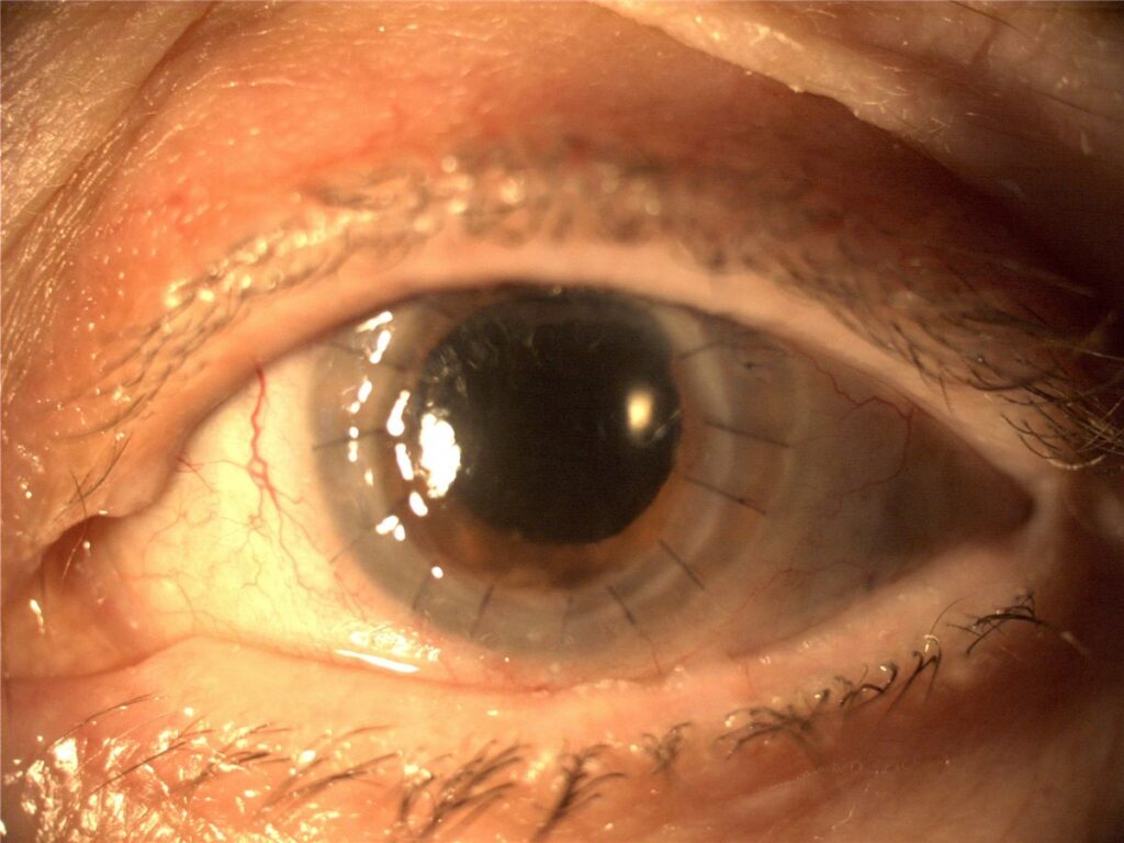
- A circular graft-host junction with visible sutures in the early postoperative period
- A clear graft with a compact stroma when the endothelium is functioning well
- Loose, broken or vascularised sutures, which are a route to infection and a trigger for rejection
- Stromal oedema and folds in Descemet membrane if the endothelium is failing
- Neovascularisation crossing the graft-host junction, a significant risk factor for rejection
Any suture that has loosened or become vascularised should be dealt with promptly, because it is one of the commonest preventable triggers of both infection and rejection.
Graft Rejection
Recognising Rejection
- Redness, pain, photophobia and blurred vision – the classic warning symptoms patients must be taught before discharge
- A Khodadoust line – a linear front of inflammatory cells advancing across the endothelium
- Keratic precipitates on the graft endothelium, with anterior chamber activity
- New graft oedema, often the first objective sign
- Subepithelial infiltrates or an advancing epithelial rejection line in anterior forms
Managing Rejection
- Intensive topical corticosteroids started urgently – outcome depends heavily on how quickly treatment begins
- Systemic or periocular steroids in severe episodes
- Steroid-sparing systemic immunosuppression in high-risk or repeatedly rejecting grafts
- Rejection is reversible if caught early; a graft left untreated may fail irreversibly
Every PK patient should leave the clinic knowing that a red, painful or newly blurred eye means being seen the same day, not at the next routine appointment.
Complications
Beyond rejection, the recognised complications include:
- High and irregular postoperative astigmatism, the commonest limit on visual outcome
- Wound dehiscence, since a full-thickness wound never regains normal corneal strength and remains vulnerable to trauma for life
- Suture-related infiltrates and microbial keratitis
- Steroid-induced ocular hypertension and glaucoma from prolonged topical steroid use
- Late endothelial cell loss leading to gradual graft failure years after successful surgery
- Recurrence of the original disease within the graft, particularly with corneal dystrophies
The permanent weakness of the full-thickness wound is a point worth making explicitly to patients, who should use eye protection for contact sports and manual work indefinitely.
Prognosis
Graft survival depends heavily on the indication and on the vascularity of the recipient bed.
- Keratoconus and corneal dystrophies in an avascular, uninflamed eye carry the best long-term graft survival
- Grafts placed in vascularised, inflamed or chemically injured eyes have a substantially higher rejection and failure rate
- Repeat grafts carry a poorer prognosis than primary grafts
- Endothelial cell density declines steadily after surgery, so late graft failure can occur even after years of clarity
- Visual outcome is often limited by astigmatism rather than by graft clarity itself
A clear graft and a good visual result are not the same thing, which is why refractive rehabilitation is as much a part of success as surgical technique.


Document what you see
Two smartphone imaging tools built for everyday clinic use — one for the slit lamp, one for the fundus.
From Choroida — the team behind this siteReferences
- Tan DTH, Dart JKG, Holland EJ, Kinoshita S. Corneal transplantation. The Lancet. 2012.
- Williams KA, Lowe M, Bartlett C, et al. Risk factors for human corneal graft failure within the Australian corneal graft registry. Transplantation. 2008.
- Price MO, Price FW. Endothelial keratoplasty – a review. Clinical and Experimental Ophthalmology. 2010.
- Penetrating Keratoplasty. EyeWiki, American Academy of Ophthalmology.
- Corneal Graft Rejection. StatPearls, NCBI Bookshelf.
Test yourself
A few questions straight from this article.
-
In penetrating keratoplasty, how much of the recipient cornea is replaced by donor tissue?
Penetrating keratoplasty removes a circular disc of the full-thickness cornea and replaces it with a matching donor button secured by sutures. -
Which corneal transplant technique replaces Descemet membrane and the endothelium alone?
DMEK transplants Descemet membrane with its endothelium alone, whereas DSAEK also carries posterior stroma and DALK replaces stroma while keeping the host endothelium. -
Which question most decides between penetrating keratoplasty and a lamellar graft at planning?
If the host endothelium is healthy, a lamellar procedure is almost always the better operation; full-thickness surgery is reserved for full-thickness pathology. -
Why is systemic immunosuppression not routinely required after corneal transplantation?
The avascular, immune-privileged cornea gives graft survival that compares favourably with solid organ transplantation without routine systemic immunosuppression. -
The open-sky stage of penetrating keratoplasty carries which specific intraoperative risk?
Because the globe is fully open at trephination, penetrating keratoplasty risks suprachoroidal haemorrhage and expulsion, which is why lamellar surgery is safer. -
A Khodadoust line after penetrating keratoplasty represents which finding?
The Khodadoust line is a linear front of inflammatory cells advancing across the graft endothelium and is a hallmark of endothelial graft rejection. -
Which finding is often the first objective sign of corneal graft rejection after keratoplasty?
New graft oedema is often the first objective sign of rejection, appearing alongside keratic precipitates and anterior chamber activity. -
What is the urgent first-line treatment for an acute corneal graft rejection episode?
Rejection is reversible if caught early, and the outcome depends heavily on how quickly intensive topical corticosteroids are started. -
Which complication most commonly limits the visual result after penetrating keratoplasty?
Astigmatism, not graft clarity, is the commonest limit on visual outcome, and many patients need rigid gas-permeable lenses for best vision. -
Which situation carries the best long-term graft survival after penetrating keratoplasty?
Keratoconus and corneal dystrophies in an avascular, uninflamed eye carry the best long-term survival, while vascularised or repeat grafts fare substantially worse.

Writes and reviews the clinical articles on Educate. Every post is checked against peer-reviewed sources before it goes live.


