
Essential blepharospasm is a focal dystonia causing involuntary, bilateral, forceful contraction of the orbicularis oculi muscles, ranging from frequent excessive blinking in mild disease to sustained, functionally blinding eyelid closure in severe cases.
It is a neurologic movement disorder, not a primary eye disease, though it is ophthalmologists who most often make the diagnosis, because patients typically present describing an eye problem rather than recognizing the condition as neurologic in origin.
It is more common in women than men and typically emerges in mid-to-late adulthood, and because its early features (frequent blinking, mild eyelid twitching) so closely resemble ordinary ocular surface irritation, patients often see several practitioners and try multiple courses of dry eye treatment before the underlying dystonic diagnosis is recognized.
Pathogenesis
Essential blepharospasm is thought to arise from dysfunction within the basal ganglia and its connections, disrupting the normal central regulation of blink and eyelid closure, rather than from any abnormality of the orbicularis muscle or the facial nerve supplying it.
The exact mechanism remains incompletely understood, and the condition is generally considered idiopathic in the great majority of patients, though it shares underlying pathophysiologic features with other focal dystonias affecting different body regions.
Clinical Presentation
The disease typically begins subtly, in the fifth to seventh decade of life, with increased blink frequency and mild eyelid twitching that gradually progresses over months to years into more sustained, forceful spasms.
As the condition advances, spasms can become severe and prolonged enough to functionally close the eyes for extended periods, impairing a patient’s ability to drive, read, or navigate their environment safely, despite the eyes themselves — and vision, when the lids are open — being entirely normal.
Some patients describe a functional, though not organic, blindness during the most severe spasm episodes, since the eyelids are forced shut regardless of the patient’s intent, and this functional impairment — despite a completely normal eye exam — is part of what makes the condition so disruptive to daily activities like driving.
All-fit Slit-Lamp Adapter
Record and share exactly what you see at the slit lamp. One adapter fits any slit lamp or surgical microscope — and any smartphone.
From Choroida — the team behind this siteTriggers and Modulating Factors
- Bright light, a common and often prominent trigger
- Stress and fatigue
- Reading, watching television, or other sustained visual tasks
- Wind
- Certain “sensory tricks” (touching the brow, chewing gum, humming, or other specific maneuvers) can transiently suppress spasms in some patients — a phenomenon seen across dystonias generally and a useful supportive clue when present
Exam Findings
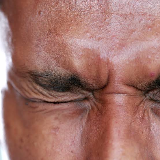
- Bilateral, synchronous, involuntary contractions of the orbicularis oculi, ranging from frequent blinking to sustained forceful closure
- A completely normal ocular exam otherwise — normal ocular surface, normal vision when the eyes are open, no structural eyelid abnormality causing the spasms
- Absence of spasms during sleep, a feature shared with most dystonias and useful in distinguishing blepharospasm from some other causes of abnormal eyelid movement
- In more advanced or associated cases, spasm involving other facial muscles (Meige syndrome, when blepharospasm occurs together with oromandibular dystonia)
Differential Diagnosis
- Hemifacial spasm — unilateral, involves lower facial muscles in addition to the eyelid, and persists during sleep, unlike essential blepharospasm; caused by vascular compression of the facial nerve rather than being a primary dystonia
- Secondary blepharospasm from ocular surface irritation — dry eye, blepharitis, or another surface condition driving reflexive, protective squeezing; addressing the underlying surface disease resolves the spasm, distinguishing it from primary essential blepharospasm
- Tardive dyskinesia — related to antipsychotic medication exposure, typically with other orofacial dyskinetic movements beyond just the eyelids
- Floppy eyelid syndrome and other causes of chronic ocular irritation, which can produce secondary blinking or squeezing that should be distinguished from a primary dystonic process
Ruling out an ocular surface cause is an important early step, because treating underlying dry eye or blepharitis is far simpler than managing primary blepharospasm.
A genuine surface trigger, when present and corrected, resolves the associated squeezing without any need for the treatments discussed below.
Diagnostic Evaluation
The diagnosis is made clinically, based on the characteristic pattern of bilateral, involuntary, sleep-sparing orbicularis spasm occurring in an eye that is otherwise structurally and functionally normal when the lids are open.
A careful ocular surface exam is essential before attributing the presentation to primary dystonia, precisely because secondary blepharospasm from dry eye or blepharitis is common and treatable through an entirely different, simpler pathway.
No specific laboratory or imaging test confirms essential blepharospasm; the diagnosis rests on recognizing the clinical pattern and excluding the secondary causes and mimics discussed above, with neurology referral considered when other dystonic or movement disorder features are present alongside the eyelid findings.
Management
Botulinum toxin injection into the orbicularis oculi muscle is the mainstay of treatment and is effective in the substantial majority of patients, though its effect is temporary, typically lasting around three months, requiring repeated injections indefinitely to maintain control.
Injection technique and dosing are individualized based on spasm severity and distribution.
Most patients find an acceptable balance between symptom control and the mild ptosis or dry eye that can occur as a side effect of treatment near the therapeutic threshold.
Oral medications (including various anticholinergic, benzodiazepine, or other centrally acting agents) have been tried but generally provide more modest benefit than botulinum toxin and are usually reserved as adjuncts for patients with an inadequate response to injections alone.
For severe, medication-refractory disease significantly impairing function, surgical options — myectomy (removal of the orbicularis muscle) or, in select cases, procedures targeting the facial nerve — are available, generally reserved for patients who have not achieved adequate control with botulinum toxin over an extended trial.
Because the condition is chronic and lifelong, ongoing coordination between the treating ophthalmologist and the patient is central to long-term management, with injection intervals and dosing adjusted over time as the pattern and severity of spasms evolve.


Document what you see
Two smartphone imaging tools built for everyday clinic use — one for the slit lamp, one for the fundus.
From Choroida — the team behind this siteReferences
- Hallett M, Evinger C, Jankovic J, Stacy M. Update on blepharospasm: report from the BEBRF International Workshop. Neurology.
- Jankovic J, Kenney C, Grafe S, et al. Relationship between various clinical outcome assessments in patients with blepharospasm. Movement Disorders.
- American Academy of Ophthalmology. Basic and Clinical Science Course, Section 7: Orbit, Eyelids, and Lacrimal System.
Test yourself
A few questions straight from this article.
-
How is essential blepharospasm best classified?
It is a focal dystonia producing involuntary, bilateral, forceful orbicularis contraction — a neurologic movement disorder rather than a primary eye disease, even though ophthalmologists usually make the diagnosis. -
Where does the dysfunction underlying essential blepharospasm arise?
Disrupted central regulation of blink and eyelid closure arises from basal ganglia dysfunction, not from any abnormality of the orbicularis muscle or of the facial nerve supplying it. -
In which decades of life does essential blepharospasm typically begin?
Onset is subtle in the fifth to seventh decade, with increased blink frequency and mild twitching that progresses over months to years into sustained forceful spasms. -
What effect do sensory tricks such as touching the brow have in essential blepharospasm?
Manoeuvres like touching the brow, chewing gum or humming can briefly suppress spasms. This phenomenon is seen across dystonias generally and is a useful supportive clue when present. -
Which feature identifies hemifacial spasm rather than essential blepharospasm?
Hemifacial spasm is unilateral, involves lower facial muscles as well as the eyelid, and persists in sleep. It results from vascular compression of the facial nerve rather than being a primary dystonia. -
Why must ocular surface disease be excluded before diagnosing essential blepharospasm?
Dry eye or blepharitis can drive reflexive protective squeezing. Correcting a genuine surface trigger resolves the spasm entirely, which is far simpler than managing primary blepharospasm. -
What defines Meige syndrome in a patient with blepharospasm?
In more advanced or associated cases the spasm involves other facial muscles; when blepharospasm occurs together with oromandibular dystonia the combination is termed Meige syndrome. -
How is the diagnosis of essential blepharospasm established?
No laboratory or imaging test confirms the condition. Diagnosis rests on the characteristic bilateral, sleep-sparing orbicularis spasm in an otherwise normal eye, with secondary causes excluded. -
What is the mainstay of treatment for essential blepharospasm?
Botulinum toxin is effective in the substantial majority of patients, but its effect is temporary — typically lasting around three months — so injections must be repeated indefinitely to maintain control. -
For which patients with essential blepharospasm is myectomy reserved?
Surgical options such as orbicularis myectomy, or procedures targeting the facial nerve, are generally reserved for severe, refractory disease after an extended trial of botulinum toxin has failed.
