
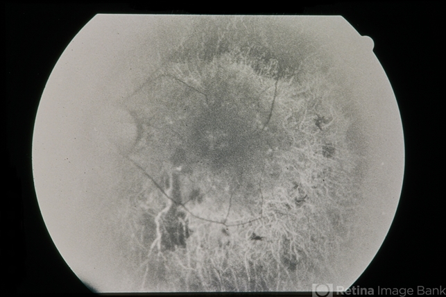
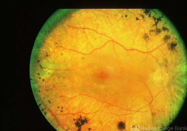
Retinitis pigmentosa (RP) is a heterogeneous group of inherited retinal disorders characterized by diffuse progressive dysfunction of predominantly rod photoreceptors with subsequent degeneration of cone photoreceptors and the retinal pigment epithelium.

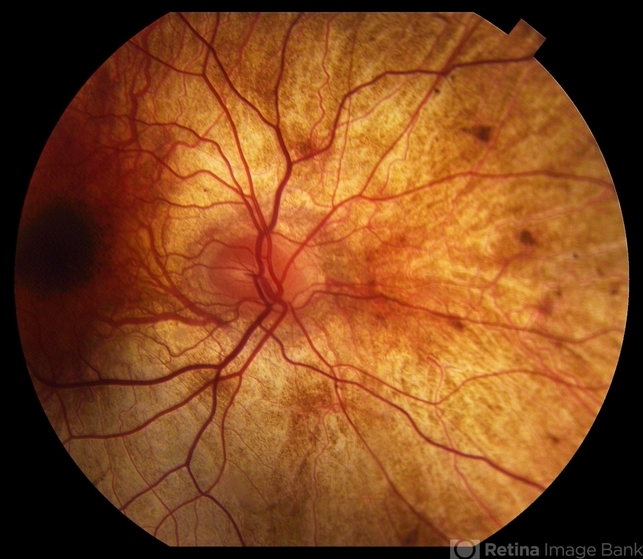
Visual impairment usually manifests as night blindness and progressive visual field loss. Retinitis pigmentosa (RP) may be seen in isolation (typical RP) or in association with systemic disease.
General treatment
Many treatments have been explored without proven benefit for the isolated forms of Retinitis pigmentosa (RP).
These include various vitamins and minerals, vasodilators, tissue therapy with placental extract, cortisone, cervical sympathectomy, injections of a hydrolysate of yeast RNA, ultrasound, transfer factor, dimethyl sulfoxide, ozone, muscle transplants, and subretinal injections of fetal retinal cells.
None of the above treatments were conducted in randomized, controlled clinical trials. It is important to note that anecdotal treatment with a subjective improvement of the visual function should be interpreted with caution due to fluctuation in visual acuity and visual fields in this disease.
ERG is a better objective measure of remaining retinal function. Any potential therapy will likely require several years of follow-up to assess efficacy due to the nature of the slow progression of this disease.
Medical therapy
Controversies surround the use of high-dose vitamin A, docosahexaenoic acid (DHA), and lutein to slow the progression of Retinitis Pigmentosa (RP).
Berson et al. conducted three large randomized trials involving 601 adult patients, assessing outcomes such as the 30-Hz cone flicker ERG.
Results indicated that higher doses of vitamin A palmitate exhibited the slowest decline in ERG amplitude, contrasting with high-dose vitamin E, which showed the fastest decline.
DHA supplementation, while not impacting RP over a 4-year interval, did show potential benefits for those starting vitamin A for the first time, slowing visual field and ERG amplitude loss in initial years.
In another study, lutein (12 mg/day) combined with high-dose vitamin A and DHA showed mixed results. While the rate of decline in the total point score for the HFA 30-2 program remained unaffected, a significant effect was observed on the rate of decline for the HFA 60-4 total point score.
Despite these findings, debates persist regarding the interpretation of study outcomes, emphasizing the need for cautious consideration of high-dose vitamin A and supplements due to potential side effects.
Treatment Considerations and Follow-up
For patients developing cystic macular lesions (about 30%), potential benefits have been identified with oral acetazolamide, topical dorzolamide or brinzolamide drops, and intravitreal steroids in some cases.
Intravitreal anti-VEGF injections have also demonstrated effectiveness in select cases. Light deprivation has not proven beneficial, but the use of ultraviolet and short-wavelength blocking sunglasses for outdoor activities is generally advised.
Annual ocular examinations are typically sufficient for measuring visual acuity and Goldmann’s visual field. However, initiation of medical treatment may necessitate more frequent visits and laboratory blood work.
Annual assessments of vitamin A levels and liver function are recommended for patients undergoing treatment, considering potential complications.
Treatment of Rare Forms and Future Directions
Patients with hereditary abetalipoproteinemia and Refsum disease necessitate unique treatment approaches, emphasizing low-fat diets and restricting phytanic acid-containing foods, respectively.
Future directions in RP management include gene therapy, with adeno-associated virus (AAV) based gene transfer showing promise in animal models and humans with specific genetic mutations.
Ciliary neurotrophic factor (CNTF) has demonstrated the ability to slow retinal degeneration in animal models, but its long-term efficacy in humans remains inconclusive.
Retinal prostheses, such as the FDA-approved ARGUS II implant, show potential for patients with end-stage RP, while gene therapy for RPE65 mutations has received FDA approval, offering hope for improved functional vision in younger patients.
Surgical Interventions
Surgical options include the ARGUS II implant for end-stage RP patients, stimulating the retina electrically and providing subjective improvements in mobility.
The Orion implant, focusing on electrodes placed into the visual cortex of the brain, is currently under investigation. Notably, while the ARGUS II implant has shown positive outcomes, the manufacturing has been discontinued, redirecting efforts toward the Orion implant.
If cataracts develop, surgical removal is generally deferred until the patient’s ability to read with a better eye diminishes. Cataract surgery has demonstrated significant improvements in visual acuity for RP patients.
Complications and Safety Measures
Toxicity from vitamin A treatment is rare but requires monitoring, with pretreatment assessments and annual checks of fasting serum vitamin A levels and liver function.
High doses of vitamin A should be avoided during pregnancy due to potential birth defects. Long-term supplementation in older adults may impact bone density, necessitating consultation for postmenopausal women and men over 49.

Patients with renal issues or on chronic doxycycline should avoid vitamin A supplementation due to potential complications.
The 5-year study of the ARGUS II implant supports its long-term safety and benefits, with complications such as conjunctival erosion and hypotony being rare but not impossible, emphasizing the need for vigilant monitoring.
As surgical and medical approaches evolve, optimizing patient outcomes remains a collaborative effort requiring ongoing research and careful consideration of individual patient needs.
Would you have interest in taking retinal images with your smartphone?
Fundus photography is superior to fundus analysis as it enables intraocular pathologies to be photo-captured and encrypted information to be shared with colleagues and patients.
Recent technologies allow smartphone-based attachments and integrated lens adaptors to transform the smartphone into a portable fundus camera and Retinal imaging by smartphone.
RETINAL IMAGING BY YOUR SMARTPHONE
References
- da Cruz L, Coley BF, Dorn J, Merlini F, Filley E, Christopher P, Chen FK, Wuyyuru V, Sahel J, Stanga P, Humayun M, Greenberg RJ, Dagnelie G; Argus II Study Group. The Argus II epiretinal prosthesis system allows letter and word reading and long-term function in patients with profound vision loss. Br J Ophthalmol 2013 May;97(5):632-6.
- Luo YH, Zhong JJ, da Cruz L. The use of Argus® II retinal prosthesis by blind subjects to achieve localisation and prehension of objects in 3-dimensional space. Graefes Arch Clin Exp Ophthalmol. 2014 Dec 31. [Epub ahead of print].
- Stronks HC, Dagnelie G. The functional performance of the Argus II retinal prosthesis. Expert Rev Med Devices. 2014 Jan;11(1):23-30..
- Bastek JV, Heckenlively JR, Straatsma BR. Cataract surgery in retinitis pigmentosa patients. Ophthalmology 1982;89(8):880-884.
- Feskanich D, Singh V, Willett WC, Colditz GA: Vitamin A intake and hip fractures among postmenopausal women. JAMA. 2002;287(1):47-54.
- Michaelsson K, Lithell H, Vessby B, Melhus H: Serum retinol levels and the risk of fracture. N Engl J Med. 2003;348(4):287-294.
- deCruz et al. Five-Year Safety and Performance Results from the Argus II Retinal Prosthesis. Ophthalmology. 2016 Oct;123(10):2248-54.
