
Tamoxifen Retinopathy is the kind of diagnosis that hides in plain sight at the macula.
Tiny refractile crystals sit quietly around the fovea of a woman being treated for breast cancer and are easily dismissed as an incidental speckle or hard drusen.
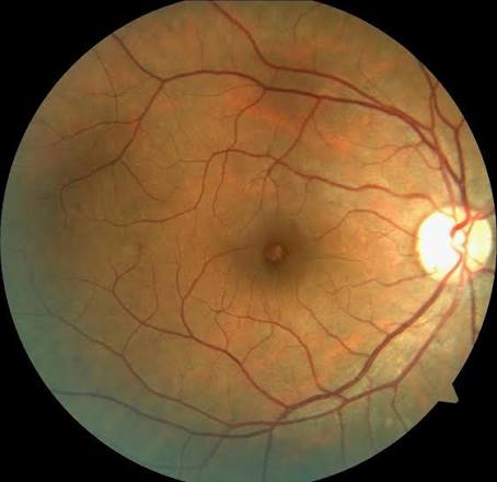
Yet those crystals, and the foveal cavitation that can accompany them, are a recognised sign of drug toxicity.
For clinicians, recognizing Tamoxifen Retinopathy matters because it changes the conversation with the oncologist and, occasionally, the choice of drug.
A careful macular examination and an OCT in any patient on long-term tamoxifen is what turns a missed speckle into a documented diagnosis.
What Is Tamoxifen Retinopathy?
Tamoxifen Retinopathy is a drug-induced crystalline maculopathy caused by the selective oestrogen receptor modulator tamoxifen, used mainly for oestrogen-receptor-positive breast cancer.
Its defining features are:
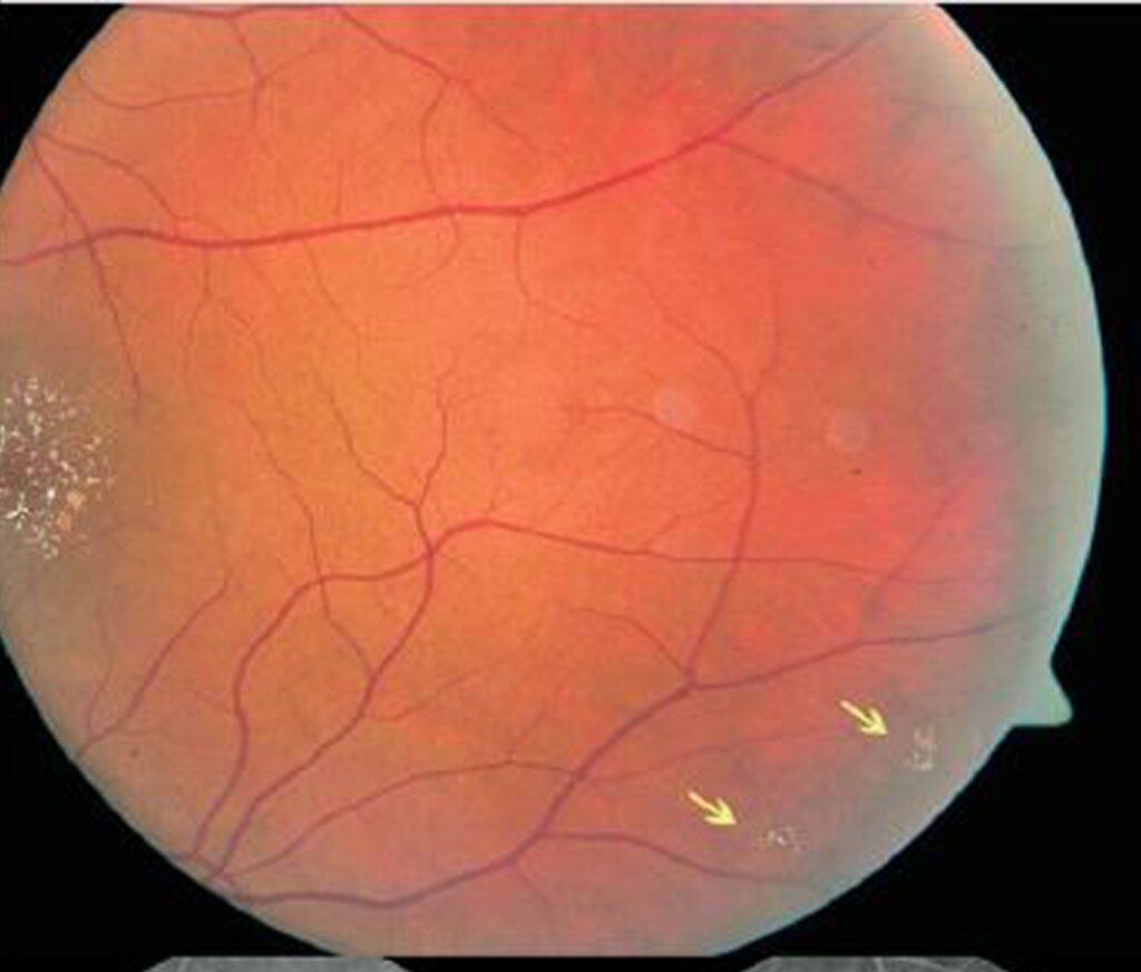
- Refractile, yellow-white crystalline deposits in the inner retina around the fovea
- Usually bilateral and roughly symmetric
- In more advanced disease, foveal cystoid or cavitary spaces on OCT
- Variable macular pigmentary change and, sometimes, macular oedema
It is best thought of as a spectrum, from a few asymptomatic crystals to a symptomatic cavitary maculopathy.
Epidemiology and Dose
The risk is tied to how much drug the retina has seen.
- Classic, florid retinopathy was first described with the very high cumulative doses used in early regimens
- Crystalline deposits and foveal cavitation are now recognised even at conventional low doses (around 20 mg/day) taken for years
- Long duration and high cumulative dose are the main risk factors
- Most patients are middle-aged or older women on adjuvant therapy
Because low-dose disease is real, no patient on long-term tamoxifen is automatically “too low a dose” to be affected.
Fundus Explorer Pro
Photograph the retinal findings described here with the phone already in your pocket — 22 D optics and built-in illumination in one handheld unit.
From Choroida — the team behind this sitePathophysiology
The crystals are thought to be a marker of retinal injury rather than deposits of the drug itself.
- They are believed to represent products of axonal and neuronal degeneration in the inner retinal layers
- Tamoxifen is lipophilic and accumulates in tissues, which fits its slow, cumulative retinal effect
- The exact molecular mechanism remains incompletely understood
Seeing the crystals as a footprint of inner-retinal degeneration explains why they can persist long after the drug is stopped.
Clinical Presentation
Symptoms
Most patients notice little or nothing.
- Often entirely asymptomatic and found on routine examination
- Reduced vision, metamorphopsia or colour disturbance when the fovea is involved
- Symptoms are usually mild relative to the striking appearance
Examination Findings
The macular picture is characteristic once looked for.
- Refractile yellow-white crystals clustered in the inner retina around the fovea
- Bilateral, broadly symmetric distribution
- Occasional macular pigmentary mottling or oedema
- A quiet peripheral retina and no vitritis
Fine refractile macular crystals in a patient on tamoxifen should prompt the diagnosis until proven otherwise.
Why Tamoxifen Retinopathy Is So Often Missed
Several factors conspire to keep the diagnosis hidden.
- The crystals are tiny and easily mistaken for hard drusen or an incidental finding
- Many patients are asymptomatic, so no one looks closely
- The drug history is not always volunteered or asked for
- The foveal cavitation on OCT is misread as a lamellar macular hole or as macular telangiectasia type 2
Asking about long-term tamoxifen, and reaching for the OCT, is what stops the diagnosis from slipping through.
Diagnostic Evaluation
The diagnosis is clinical, confirmed and characterised by imaging.
Optical Coherence Tomography
- Hyperreflective dots corresponding to the inner-retinal crystals
- Foveal cystoid or cavitary spaces that can mimic a macular hole or macular telangiectasia type 2
- These cavities are typically non-leaking, unlike true cystoid macular oedema
Fundus Autofluorescence and Angiography
- Autofluorescence documents associated RPE changes
- Fluorescein angiography helps separate genuine leaking oedema from the non-leaking foveal cavitation of tamoxifen
OCT is the single most useful test, both to confirm the crystals and to characterise the foveal cavitation.
Differential Diagnosis
The differential is essentially the other crystalline and foveal-cavitary maculopathies:
- Macular telangiectasia type 2 — foveal cavitation with right-angle venules and telangiectatic vessels, no drug history
- Bietti crystalline dystrophy — inherited, with crystals plus choroidal and RPE atrophy
- Ocular cystinosis — corneal and retinal crystals in a systemic metabolic context
- Other crystalline retinopathies — canthaxanthine, methoxyflurane oxalosis, talc (intravenous drug use) and nitrofurantoin
- Hard drusen — deeper, non-refractile, without foveal cavitation
The drug history and the bilateral perifoveal crystals are what point specifically to tamoxifen.
Screening and Management
Management is a shared decision, not an ophthalmic one alone.
- A baseline eye examination and periodic macular review with OCT are reasonable for patients on long-term therapy
- Findings should be discussed with the treating oncologist, as the oncological benefit of tamoxifen is substantial
- Stopping or switching the drug (for example to an aromatase inhibitor) is considered when there is significant, vision-threatening maculopathy
- The crystals often persist after cessation, whereas the foveal cystoid changes may partly improve
The ophthalmologist documents and quantifies the toxicity; the decision to continue or change treatment is made with oncology.
Prognosis
For most patients the visual outlook is good.
- Vision is usually preserved, especially with the crystals-only, non-foveal form
- Significant visual loss is uncommon at conventional adjuvant doses
- Foveal cavitation involving the centre carries a more guarded visual prognosis
In practice, the value of recognising Tamoxifen Retinopathy is early documentation and a timely conversation, not alarm.


Document what you see
Two smartphone imaging tools built for everyday clinic use — one for the slit lamp, one for the fundus.
From Choroida — the team behind this siteReferences
- Kaiser-Kupfer MI, Lippman ME. “Tamoxifen Retinopathy.” Cancer Treatment Reports. 1978;62(3):315–320.
- Pavlidis NA, Petris C, Briassoulis E, et al. “Clear Evidence That Long-Term, Low-Dose Tamoxifen Treatment Can Induce Ocular Toxicity.” Cancer. 1992;69(12):2961–2964.
- Gualino V, Cohen SY, Delyfer MN, et al. “Optical Coherence Tomography Findings in Tamoxifen Retinopathy.” American Journal of Ophthalmology. 2005;140(4):757–758.
- Doshi RR, Fortun JA, Kim BT, Dubovy SR, Rosenfeld PJ. “Pseudocystic Foveal Cavitation in Tamoxifen Retinopathy.” American Journal of Ophthalmology. 2014;157(6):1291–1298.
- American Academy of Ophthalmology. “Tamoxifen Retinopathy.” EyeWiki.
Test yourself
A few questions straight from this article.
-
Tamoxifen, the drug responsible for tamoxifen retinopathy, belongs to which class?
Tamoxifen is a selective oestrogen receptor modulator used mainly for oestrogen-receptor-positive breast cancer, and its crystalline maculopathy is a recognised drug toxicity. -
Where do the refractile crystalline deposits of tamoxifen retinopathy characteristically sit?
The defining lesion is refractile yellow-white crystalline deposits in the inner retina around the fovea, usually bilateral and roughly symmetric. -
What are the crystals of tamoxifen retinopathy currently believed to represent?
The crystals are thought to be a marker of retinal injury rather than deposits of the drug itself, representing products of axonal and neuronal degeneration in the inner retinal layers. -
At roughly what conventional daily tamoxifen dose is crystalline retinopathy now recognised?
Although florid retinopathy was first described with very high cumulative doses, crystalline deposits and foveal cavitation are now recognised at conventional low doses of around 20 mg/day taken over years. -
How do the foveal cavitary spaces of tamoxifen retinopathy behave on fluorescein angiography?
The foveal cystoid or cavitary spaces are typically non-leaking, which is how angiography separates them from genuine cystoid macular oedema. -
Which finding favours macular telangiectasia type 2 over tamoxifen retinopathy in foveal cavitation?
Macular telangiectasia type 2 produces foveal cavitation accompanied by right-angle venules and telangiectatic vessels, and there is no tamoxifen drug history. -
Which crystalline retinopathy is inherited and shows crystals with choroidal and RPE atrophy?
Bietti crystalline dystrophy is inherited and combines retinal crystals with choroidal and retinal pigment epithelial atrophy, unlike the drug-induced picture of tamoxifen. -
When is stopping or switching tamoxifen considered in a patient with crystalline maculopathy?
Because the oncological benefit of tamoxifen is substantial, stopping or switching the drug is reserved for significant vision-threatening maculopathy and is decided together with the treating oncologist. -
What typically happens to the macular findings of tamoxifen retinopathy after the drug is stopped?
The crystals often persist after cessation, in keeping with their being a footprint of inner-retinal degeneration, whereas the foveal cystoid changes may partly improve. -
Which form of tamoxifen retinopathy carries the more guarded visual prognosis?
Vision is usually preserved in the crystals-only, non-foveal form, whereas foveal cavitation involving the centre carries a more guarded visual prognosis.
