
Cone-rod dystrophy is an inherited retinal degeneration in which cone photoreceptor dysfunction predominates and typically appears first, followed later by rod involvement — the exact reverse sequence from retinitis pigmentosa, where rod dysfunction (with its characteristic early night blindness) comes first, and cone involvement follows only later in the disease course.
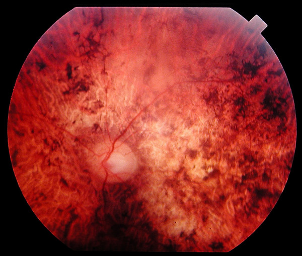
This inverted order of photoreceptor involvement is the defining clinical and electrophysiologic feature that separates the two conditions, and getting the distinction right matters directly for prognosis and genetic counseling.
Why the Order of Involvement Matters
Because cones are concentrated in and around the fovea and are responsible for central vision, color perception, and visual acuity under normal lighting conditions, cone-predominant dysfunction produces early loss of central vision, color vision abnormalities, and photophobia, often while peripheral vision and night vision remain relatively preserved for a considerable time.
This is the opposite functional pattern from retinitis pigmentosa’s classic early night blindness and peripheral field loss with preserved central vision until much later in the disease course, and recognizing which pattern a patient shows is central to correctly distinguishing the two conditions.
Clinical Presentation
- Progressive, bilateral loss of central visual acuity, often the earliest and most bothersome symptom
- Color vision abnormalities, sometimes an early or even presenting complaint, particularly for red-green discrimination though the specific pattern varies
- Photophobia — a genuinely characteristic feature, related to the predominant cone dysfunction, and often prominent enough that patients specifically avoid bright environments
- Reduced contrast sensitivity, sometimes out of proportion to the degree of visual acuity loss on standard testing
- Night vision and peripheral field are relatively preserved early in the disease course, though rod involvement and corresponding peripheral field loss and night vision difficulty do eventually develop as the disease progresses
Fundus Explorer Pro
Photograph the retinal findings described here with the phone already in your pocket — 22 D optics and built-in illumination in one handheld unit.
From Choroida — the team behind this siteExam and Testing Findings
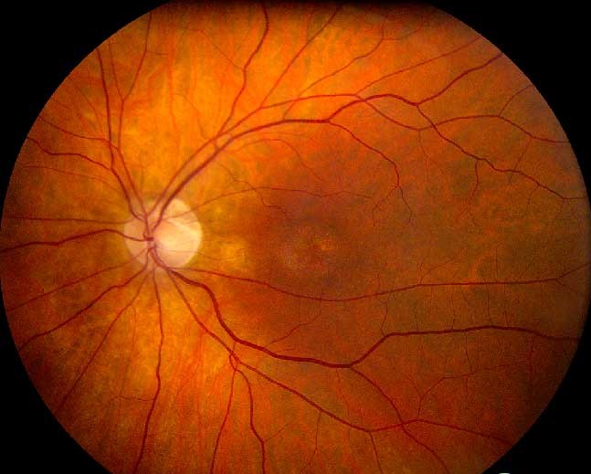
- Macular atrophy or a “bull’s eye” pattern of retinal pigment epithelial change centered on the fovea in many, though not all, cases — a finding overlapping in appearance with other causes of bull’s eye maculopathy, including hydroxychloroquine toxicity, and requiring careful correlation with medication history and the rest of the clinical picture to distinguish an inherited from an acquired cause
- Vascular attenuation and optic disc pallor developing later in the disease course, as the condition progresses
- Electroretinography showing predominant, disproportionate reduction in cone-mediated (photopic) responses relative to rod-mediated (scotopic) responses, at least in earlier stages — the specific electrophysiologic signature that most definitively distinguishes cone-rod dystrophy from retinitis pigmentosa’s opposite pattern
- Progressive field loss, developing centrally first and eventually extending peripherally as rod function also becomes affected in later disease
Genetics
Cone-rod dystrophy is genetically heterogeneous, with autosomal dominant, autosomal recessive, and X-linked inheritance patterns all described across different implicated genes (including ABCA4, also implicated in Stargardt disease, reflecting a genuine overlap and shared genetic spectrum between these related macular and retinal dystrophies).
The specific gene and inheritance pattern influence both the expected disease course and family counseling regarding recurrence risk, and increasingly determine eligibility for specific gene-targeted research and treatment programs as they become available.
Differential Diagnosis
- Retinitis pigmentosa — rod-cone (rather than cone-rod) pattern, with early night blindness and peripheral field loss preceding central vision loss, discussed in its own dedicated article on this site; the ERG pattern is the definitive way to distinguish the two when the clinical history alone leaves ambiguity
- Stargardt disease — genetically overlapping in some cases (shared ABCA4 involvement) and can show a similar macular atrophy pattern, though Stargardt disease classically presents earlier, in childhood or adolescence, and has its own characteristic flecks and “dark choroid” sign on fluorescein angiography, discussed in its own dedicated article on this site
- Achromatopsia — congenital, stationary (non-progressive) cone dysfunction from birth, rather than the progressive, later-onset pattern typical of cone-rod dystrophy, discussed in its own dedicated article on this site
- Hydroxychloroquine or other toxic maculopathy — an acquired rather than inherited cause of a similar bull’s eye maculopathy appearance, distinguished by medication history, discussed in this site’s coverage of hydroxychloroquine toxicity
Diagnostic Evaluation
Electroretinography is the essential test for confirming the cone-predominant pattern of dysfunction and distinguishing cone-rod dystrophy from retinitis pigmentosa.
OCT and fundus autofluorescence document the extent of macular atrophy and RPE change, providing an objective anatomic complement to the functional information gathered from ERG testing.
Genetic testing increasingly plays a role both in confirming the diagnosis and in identifying the specific causative gene, which is relevant for prognosis, family counseling, and, for a growing number of specific genetic subtypes, potential eligibility for gene-specific clinical trials or emerging therapies.
Management
There is no treatment that halts the underlying progressive photoreceptor degeneration for most genetic subtypes of cone-rod dystrophy; management is supportive: photophobia management with tinted lenses, low vision aids and rehabilitation as central vision declines, and genetic counseling for the family.
Regular monitoring tracks disease progression, including serial ERG, OCT, and autofluorescence imaging to document the pace of change over time.
Patients should be kept informed about the rapidly evolving landscape of gene-specific and other emerging retinal therapies, because a condition without a targeted treatment today may have one within a relevant timeframe given the pace of development in this field, for well-characterized genetic subtypes.


Document what you see
Two smartphone imaging tools built for everyday clinic use — one for the slit lamp, one for the fundus.
From Choroida — the team behind this siteReferences
- Hamel CP. Cone rod dystrophies. Orphanet Journal of Rare Diseases.
- Michaelides M, Hardcastle AJ, Hunt DM, Moore AT. Progressive cone and cone-rod dystrophies: phenotypes and underlying molecular genetic basis. Survey of Ophthalmology.
- American Academy of Ophthalmology. Basic and Clinical Science Course, Section 12: Retina and Vitreous.
Test yourself
A few questions straight from this article.
-
Cone-rod dystrophy is defined by which sequence of photoreceptor involvement?
Cone dysfunction predominates and typically appears first, with rod involvement following later. This is the exact reverse of retinitis pigmentosa, where rods go first. -
Which symptom pattern is expected early in the course of cone-rod dystrophy?
Because cones drive central vision and colour perception, early disease produces central acuity loss, colour abnormalities and photophobia, while night and peripheral vision stay relatively intact. -
Why does cone-predominant dysfunction cause early central visual loss in cone-rod dystrophy?
Cones cluster at and around the fovea and carry central acuity and colour vision, so cone-predominant degeneration strikes central vision first. -
Which macular appearance is commonly described on fundus examination in cone-rod dystrophy?
Many cases show macular atrophy or a bull's eye pattern of retinal pigment epithelial change centred on the fovea, though this is not present in every case. -
Which electroretinography pattern most definitively identifies cone-rod dystrophy?
Disproportionate reduction of cone-mediated photopic responses relative to rod-mediated scotopic responses is the electrophysiologic signature separating cone-rod dystrophy from retinitis pigmentosa. -
How does visual field loss typically progress in cone-rod dystrophy?
Field loss develops centrally first, then spreads peripherally as rod function also becomes affected in later disease. -
Which gene implicated in cone-rod dystrophy is also implicated in Stargardt disease?
ABCA4 is among the genes implicated in cone-rod dystrophy and is also implicated in Stargardt disease, reflecting a shared genetic spectrum between these dystrophies. -
Which finding distinguishes Stargardt disease from cone-rod dystrophy?
Stargardt disease has characteristic flecks and a dark choroid sign on fluorescein angiography, and classically presents earlier, in childhood or adolescence. -
Which feature distinguishes achromatopsia from cone-rod dystrophy?
Achromatopsia is congenital and non-progressive cone dysfunction from birth, unlike the progressive, later-onset pattern typical of cone-rod dystrophy. -
What does management of cone-rod dystrophy consist of for most genetic subtypes?
No treatment halts the degeneration for most subtypes, so care is supportive: tinted lenses for photophobia, low vision rehabilitation, and genetic counselling, with serial ERG, OCT and autofluorescence monitoring.
