
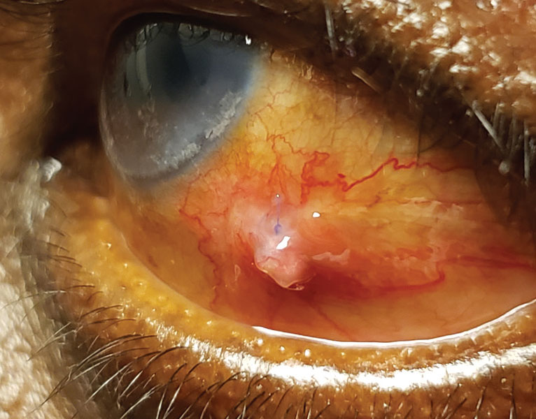
A conjunctival granuloma describes a histologic pattern, chronic granulomatous inflammation, rather than a single disease, and the real clinical work when this finding is identified lies entirely in tracing it back to its specific cause, since the range of possible underlying conditions, from a simple, locally irritating chalazion to systemic sarcoidosis, is genuinely broad and carries very different implications for each.
What “Granulomatous” Means
Granulomatous inflammation represents a specific pattern of chronic immune response, characterized by collections of macrophages and other immune cells organizing around a persistent stimulus the body has been unable to fully clear or eliminate, whether that stimulus is a foreign material, certain infectious organisms, or an immune-mediated systemic process.
A conjunctival granuloma, then, is the visible, localized mass that results when this pattern of inflammation occurs within the conjunctiva, and identifying the specific underlying cause requires looking beyond the local finding itself.
Common Causes
Chalazion-Related Granuloma
A chalazion, a chronic granulomatous inflammatory reaction to retained meibomian gland secretions, can sometimes extend through or present primarily on the conjunctival surface, producing a conjunctival granulomatous mass directly related to this common eyelid condition.
Foreign Body Reaction
A retained foreign body, including suture material from prior ocular surgery, can incite a chronic granulomatous response at the site of retention, and a relevant surgical or trauma history is an important clue supporting this cause.
Pyogenic Granuloma
Despite sharing “granuloma” in its name, a pyogenic granuloma is technically a vascular proliferative lesion rather than a true granulomatous inflammatory process, but it is frequently included in the clinical differential for a fleshy conjunctival mass and is worth distinguishing given its own distinct, generally reactive and self-limited, natural history (see pyogenic granuloma for a detailed look at this related but mechanistically distinct entity)
Sarcoidosis
Conjunctival granulomas can be a manifestation of systemic sarcoidosis, sometimes presenting as a relatively accessible, biopsiable site that can support a systemic diagnosis, making conjunctival involvement a genuinely useful diagnostic opportunity when sarcoidosis is suspected based on other systemic findings
Cat Scratch Disease (Parinaud Oculoglandular Syndrome)
Conjunctival granuloma formation, together with regional (typically preauricular) lymphadenopathy, is a classic presentation of cat scratch disease affecting the eye, and a relevant history of cat exposure or scratch supports this specific diagnosis
Other Infectious and Inflammatory Causes
Tuberculosis, and other granulomatous infections, can occasionally present with conjunctival granuloma formation, generally in the context of other suggestive systemic findings or known exposure risk
All-fit Slit-Lamp Adapter
Record and share exactly what you see at the slit lamp. One adapter fits any slit lamp or surgical microscope — and any smartphone.
From Choroida — the team behind this siteDiagnostic Evaluation
- A thorough history, specifically asking about prior ocular surgery or trauma, animal exposure (particularly cats), known systemic conditions, and any associated symptoms suggesting an underlying eyelid process such as chalazion
- Careful examination of the eyelids and meibomian glands, looking for a primary chalazion that might be the source of a conjunctival extension
- Assessment for regional lymphadenopathy, particularly relevant for cat scratch disease evaluation
- Biopsy of the conjunctival lesion, with histopathologic examination, is often necessary to confirm the granulomatous nature of the lesion and can sometimes provide additional clues (such as specific staining patterns or organism identification) pointing toward a specific underlying cause
- When sarcoidosis is suspected, biopsy with appropriate staining, combined with systemic evaluation including chest imaging and serum markers, supports this specific diagnosis
Management
Treating the Underlying Cause
Management is directed entirely at the specific underlying cause once identified, whether that means addressing a primary chalazion, removing a retained foreign body, treating an underlying infection, or managing systemic sarcoidosis in coordination with the appropriate specialists.
Biopsy as Both Diagnostic and Therapeutic
For many causes of conjunctival granuloma, excisional biopsy serves a dual purpose, providing definitive histopathologic diagnosis while also removing the lesion itself, making this a genuinely efficient approach for many presentations of this finding.
Finding the cause
Ask about previous surgery, trauma, foreign bodies, and long-standing suture material, since these are the usual triggers. Examine the lesion and the surrounding conjunctiva, and evert the lids to look for hidden foreign material. If the lesion is solitary and associated with a suture, removal of the material and excision of the granuloma usually cures it. Send any removed tissue for histology, and consider tests for sarcoidosis or infection when the lesions are multiple or recurrent.
Prognosis
Prognosis depends entirely on the underlying cause, ranging from excellent with complete resolution after treatment of a straightforward chalazion-related or foreign body-related granuloma, to a prognosis tied to the course of an underlying systemic condition when sarcoidosis or another systemic process is responsible.
Given this wide range of possible underlying causes and their correspondingly different implications, a conjunctival granuloma should always prompt a deliberate, systematic search for its specific cause rather than being treated as a uniform, single diagnostic and management category.


Document what you see
Two smartphone imaging tools built for everyday clinic use — one for the slit lamp, one for the fundus.
From Choroida — the team behind this siteReferences
- Shields JA, Shields CL. Eyelid, Conjunctival, and Orbital Tumors: An Atlas and Textbook. 3rd ed. Philadelphia: Wolters Kluwer; 2016.
- Margileth AM, Wear DJ, English CK. Systemic cat scratch disease: report of 23 patients with prolonged or recurrent severe bacterial infection. J Infect Dis. 1987;155:390-402.
- Rothova A. Ocular involvement in sarcoidosis. Br J Ophthalmol. 2000;84:110-116.
- Chanda D, Brenner AV, Fullick E, et al. Conjunctival biopsy in the diagnosis of sarcoidosis. Eye (Lond). 2007;21:823-824.
