Fuchs heterochromic iridocyclitis is the great impostor of anterior uveitis.
It is a chronic, low-grade inflammation that produces almost no redness, no pain, and no synechiae—so it is frequently missed or mislabeled for years.
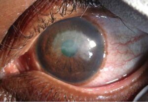
Instead of an angry eye, the clue is a quiet one with subtle iris heterochromia, a scattering of fine stellate deposits on the cornea, and an early cataract.
For clinicians, recognizing Fuchs heterochromic iridocyclitis (FHIC)—also called Fuchs uveitis syndrome—matters because treating it like ordinary uveitis with aggressive steroids does more harm than good.
The diagnosis is made at the slit lamp, and a high index of suspicion in a white, quiet eye is what prevents years of misdirected treatment.
What Is Fuchs Heterochromic Iridocyclitis?
FHIC is a chronic, usually unilateral anterior uveitis defined by a characteristic clinical triad.
The classic features are:
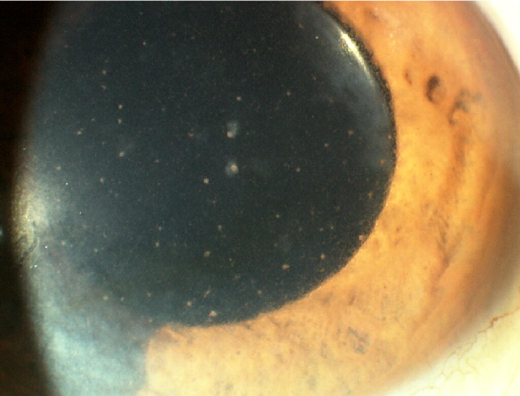
- Diffuse, small, stellate keratic precipitates over the whole corneal endothelium
- Iris heterochromia with stromal atrophy
- Early posterior subcapsular cataract
The inflammation is remarkably indolent, and the eye typically stays white and comfortable throughout.
Epidemiology
FHIC accounts for roughly 2–5% of all uveitis cases.
- Most patients present in the third or fourth decade of life
- There is no strong sex predilection
- Involvement is unilateral in the large majority of cases, though bilateral disease is occasionally seen
Because symptoms are minimal, the diagnosis is often delayed for years until a cataract or glaucoma develops.
All-fit Slit-Lamp Adapter
Record and share exactly what you see at the slit lamp. One adapter fits any slit lamp or surgical microscope — and any smartphone.
From Choroida — the team behind this sitePathogenesis
The exact cause remains debated, but the evidence increasingly points to a chronic viral trigger.
- Rubella virus is the most strongly implicated agent; aqueous samples frequently contain anti-rubella antibodies and viral genome, and the incidence of FHIC has fallen sharply in rubella-vaccinated populations
- Cytomegalovirus is detected in the aqueous of a subset of cases, especially in East Asian populations, and may explain the clinical overlap with Posner–Schlossman syndrome
- The end result is a low-grade, antibody-mediated inflammation that slowly damages the iris stroma and its vasculature
This slow immune-mediated damage—rather than an acute inflammatory storm—explains the characteristic iris atrophy and the eye’s deceptively quiet appearance.
Clinical Presentation
Symptoms
The hallmark is how little the patient complains.
- Gradual blurring of vision from cataract or vitreous opacities
- Floaters
- No pain and no redness—the eye stays white and quiet
Examination Findings
The diagnosis is essentially a slit-lamp diagnosis.
- Keratic precipitates: small, whitish, stellate or round, distributed diffusely over the entire endothelium—often with fine connecting filaments
- Iris: stromal atrophy, loss of crypts, and blunted architecture producing heterochromia
- Iris nodules: Koeppe nodules at the pupil margin or Busacca nodules in the stroma may be present
- Anterior chamber: mild cells with minimal flare
- No posterior synechiae, even after years of inflammation
- Vitreous: cells and opacities are common and account for many of the floaters
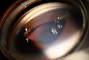
- Fine iris or angle vessels; a filiform hemorrhage from the angle on paracentesis (Amsler–Verrey sign)
Diffuse stellate keratic precipitates distributed over the whole endothelium—rather than clumped inferiorly—are the single most useful diagnostic clue.
A Note on Heterochromia
Iris colour change is classic but can be surprisingly hard to read.
- In light irides, the affected eye usually becomes hypochromic (lighter) as the stroma atrophies
- In dark irides, atrophy can expose the pigment epithelium and make the affected eye appear darker
- Heterochromia may be subtle or absent, so its absence never excludes the diagnosis
Always compare the two irides in good light before concluding there is no colour difference.
Why Fuchs Uveitis Is So Often Missed
Few conditions are misdiagnosed as consistently as Fuchs uveitis.
- The eye is white and painless, so patients present late and clinicians do not suspect uveitis
- The keratic precipitates are small and easily overlooked unless the whole endothelium is scanned
- Heterochromia is subtle, particularly in brown irides, and is frequently missed
- Many patients are treated for years with topical steroids for “idiopathic” uveitis, collecting side effects without benefit
The single most useful habit is to consider FHIC in any chronic, low-grade, unilateral uveitis that lacks synechiae.

Diagnostic Evaluation
The diagnosis is clinical, but a structured examination confirms it and excludes its mimics.
Clinical Assessment
- A careful slit-lamp survey of the entire endothelium for diffuse stellate KPs
- Comparison of both irides in daylight for subtle heterochromia
- Deliberate confirmation of the conspicuous absence of posterior synechiae
Gonioscopy
- Fine, fragile new vessels may be seen in the angle
- These vessels bleed as a filiform hemorrhage during paracentesis or surgery (Amsler–Verrey sign)
Ancillary Testing
- Aqueous PCR or antibody analysis for rubella and cytomegalovirus can confirm the diagnosis in atypical cases
- Such testing is generally reserved for diagnostic uncertainty or research rather than routine practice
Routine cases need no laboratory testing—the diagnosis lives at the slit lamp.
Differential Diagnosis
Both the uveitis and the heterochromia have important mimics:
- Posner–Schlossman syndrome — episodic IOP spikes, also CMV-associated
- Herpetic (HSV/VZV) anterior uveitis — sectoral iris atrophy and raised IOP
- Acute anterior uveitis (e.g. HLA-B27) — painful, red, with synechiae
- Other causes of heterochromia — congenital Horner syndrome, ocular siderosis, ocular melanocytosis
The combination of a white eye, diffuse stellate KPs, and absent synechiae is what separates FHIC from these conditions.
Complications
Most of the visual morbidity comes from complications, not the inflammation itself.
- Cataract — typically posterior subcapsular, and the commonest cause of gradual visual decline
- Secondary open-angle glaucoma — common, often driven by chronic trabecular inflammation, and frequently harder to control than primary open-angle glaucoma
- Vitreous opacities producing persistent floaters
- Occasionally, spontaneous hyphema from fragile angle vessels
Glaucoma, not the uveitis, is the main long-term threat to vision.
Management
Treatment is deliberately conservative.
- Mild anterior chamber activity usually needs no treatment
- Topical corticosteroids have limited benefit and add steroid-related cataract and glaucoma risk—reserve short courses for genuine symptomatic flares
- Systemic immunosuppression is not required and does not change the course of the disease
- Cataract surgery generally gives excellent results; anticipate a small hyphema from angle vessels (Amsler sign) and a slightly higher rate of postoperative inflammation
- Glaucoma is managed medically first, but frequently progresses to require trabeculectomy or a drainage device
Recognizing that FHIC does not respond to—and does not need—aggressive anti-inflammatory therapy is the key management principle.
Prognosis
The overall visual prognosis is good.
- Cataract surgery is usually successful with careful technique
- Vision is preserved in most eyes when glaucoma is detected and controlled early
- Lifelong monitoring of intraocular pressure is essential
In practice, the greatest service to the patient is making the diagnosis early, avoiding unnecessary steroids, and watching the pressure.
Would you like to document subtle findings like stellate keratic precipitates with your smartphone?


Document what you see
Two smartphone imaging tools built for everyday clinic use — one for the slit lamp, one for the fundus.
From Choroida — the team behind this siteReferences
- Jones NP. “Fuchs’ Heterochromic Uveitis: a Reappraisal of the Clinical Spectrum.” Eye. 1991;5(6):649–661.
- Chee SP, Jap A. “Presumed Fuchs Heterochromic Iridocyclitis and Posner–Schlossman Syndrome: Comparison of Cytomegalovirus-Positive and -Negative Eyes.” American Journal of Ophthalmology. 2008;146(6):883–889.
- Quentin CD, Reiber H. “Fuchs Heterochromic Cyclitis: Rubella Virus Antibodies and Genome in Aqueous Humor.” American Journal of Ophthalmology. 2004;138(1):46–54.
- Birnbaum AD, Tessler HH, Schultz KL, et al. “Epidemiologic Relationship Between Fuchs Heterochromic Iridocyclitis and the United States Rubella Vaccination Program.” American Journal of Ophthalmology. 2007;144(3):424–428.
- American Academy of Ophthalmology. “Fuchs Heterochromic Iridocyclitis.” EyeWiki.
Test yourself
A few questions straight from this article.
-
Fuchs heterochromic iridocyclitis is best characterised as which pattern of intraocular inflammation?
The inflammation is indolent and typically one-sided, so the eye stays white and comfortable rather than red and painful. -
What proportion of all uveitis cases does Fuchs heterochromic iridocyclitis account for?
Fuchs uveitis makes up only about 2-5% of uveitis, and because symptoms are minimal the diagnosis is often delayed for years. -
Which virus is most strongly implicated in the pathogenesis of Fuchs heterochromic iridocyclitis?
Aqueous samples frequently contain anti-rubella antibodies and viral genome, and the incidence has fallen sharply in rubella-vaccinated populations. -
Which slit-lamp finding is the single most useful diagnostic clue in Fuchs heterochromic iridocyclitis?
The precipitates are small, whitish and scattered across the entire endothelium rather than clumped inferiorly, often with fine connecting filaments. -
In Fuchs heterochromic iridocyclitis, which finding stays conspicuously absent despite years of inflammation?
Absence of posterior synechiae is so characteristic that it should be deliberately confirmed, and it helps separate Fuchs uveitis from other chronic anterior uveitides. -
In a patient with light-coloured irides, how does the affected iris typically change in Fuchs uveitis?
Stromal atrophy makes a light iris hypochromic, whereas in dark irides the exposed pigment epithelium can make the affected eye look darker instead. -
What is the Amsler-Verrey sign described in Fuchs heterochromic iridocyclitis?
Fine fragile new vessels in the angle bleed as a thread-like hemorrhage during paracentesis or surgery, which should be anticipated at cataract surgery. -
Which combination separates Fuchs heterochromic iridocyclitis from HLA-B27 acute anterior uveitis?
HLA-B27 disease is painful and red with synechiae, while Fuchs uveitis presents as a white eye with diffuse stellate precipitates and no synechiae. -
How should mild anterior chamber activity in Fuchs heterochromic iridocyclitis usually be managed?
Treatment is deliberately conservative: steroids add cataract and glaucoma risk with little benefit, and systemic immunosuppression does not change the disease course. -
Which complication is the main long-term threat to vision in Fuchs heterochromic iridocyclitis?
Secondary open-angle glaucoma is common and often harder to control than primary open-angle disease, so lifelong pressure monitoring is essential.

Writes and reviews the clinical articles on Educate. Every post is checked against peer-reviewed sources before it goes live.