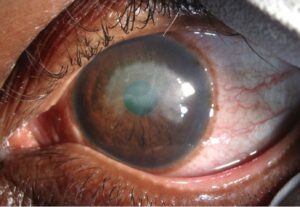
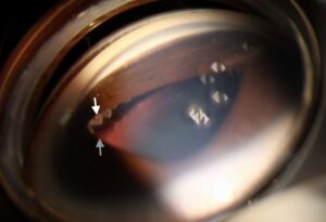
Viral anterior uveitis caused by cytomegalovirus, herpes simplex virus, or varicella zoster virus produces a recognizable combination of anterior chamber inflammation and elevated intraocular pressure that distinguishes it from most other forms of anterior uveitis.
Missing the viral cause of an anterior uveitis means treating with steroids alone, which controls the inflammation temporarily but does nothing for the infection and allows the corneal endothelium to keep losing cells.
Viral Anterior Uveitis: Why It Looks Different
HLA-B27 and autoimmune anterior uveitis rarely raise pressure this way; viral anterior uveitis commonly raises intraocular pressure during the acute attack, sometimes markedly, because of trabeculitis.
It is typically unilateral, recurrent, and low-grade to moderate in its inflammatory activity, and corneal endothelial changes are common.
Cytomegalovirus Anterior Uveitis
CMV anterior uveitis occurs in immunocompetent patients, in immunocompetent hosts as well as, and it has become increasingly recognized with the availability of aqueous PCR testing.
Features include:
- Unilateral, recurrent or chronic anterior uveitis with elevated IOP
- Coin-shaped or medium-sized keratic precipitates, sometimes with an endothelial pattern
- A clinical overlap with Posner-Schlossman syndrome (glaucomatocyclitic crisis), which many cases previously labeled idiopathic are now attributed to CMV (see Posner-Schlossman syndrome)
- Progressive endothelial cell loss over repeated attacks
Herpes Simplex and Varicella Zoster Anterior Uveitis
HSV and VZV anterior uveitis often follow or accompany keratitis, though uveitis can occur without visible corneal disease.
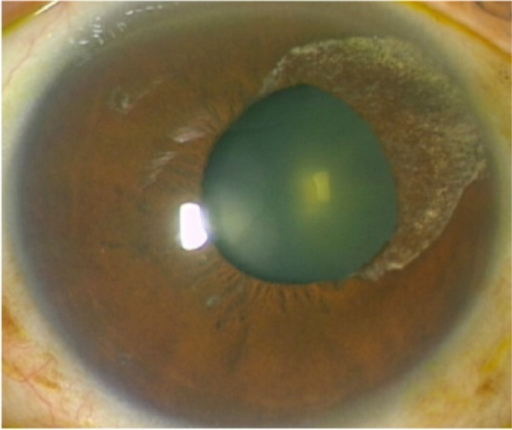
- Sectoral iris atrophy is a helpful sign, more classically associated with VZV but seen with HSV as well
- Elevated IOP during the acute attack is common
- Recurrent episodes are typical, and each attack risks further endothelial and iris damage
- A history of prior herpes zoster ophthalmicus or recurrent herpetic keratitis supports the diagnosis (see herpes zoster ophthalmicus and herpes simplex keratitis)
Diagnosis
Clinical suspicion is often enough to start empirical antiviral treatment, but aqueous humor PCR confirms the specific virus when the diagnosis is unclear, the course is atypical, or treatment fails.
Testing is particularly useful to distinguish CMV from HSV and VZV, since it changes the choice and duration of antiviral therapy.
IOP should be checked and trended at each visit, since the pressure pattern is itself a diagnostic clue.
Management
Antiviral Therapy
Oral valacyclovir or acyclovir is used for HSV and VZV disease, and oral valganciclovir or topical ganciclovir is used for CMV anterior uveitis.
Duration is typically weeks to months, and some patients with CMV need long-term low-dose suppressive therapy to prevent recurrence.
Anti-Inflammatory and Pressure Control
Topical corticosteroids control the anterior chamber inflammation, generally started alongside antiviral cover rather than alone.
IOP-lowering medications are used during acute attacks, and pressure often normalizes as inflammation resolves, though some patients develop secondary glaucoma after repeated episodes.
Long-Term Care
Recurrent attacks warrant discussion of prophylactic oral antiviral therapy, similar to the approach used for recurrent herpetic keratitis.
Endothelial cell counts should be monitored over time in patients with repeated attacks, since cumulative loss can lead to corneal decompensation.
Prognosis
Most patients maintain good vision with appropriate antiviral and anti-inflammatory treatment, though repeated attacks carry a cumulative risk to the endothelium and to IOP control.
Recognizing the viral cause changes management meaningfully, since steroid therapy alone is not enough and does not prevent recurrence.


Document what you see
Two smartphone imaging tools built for everyday clinic use — one for the slit lamp, one for the fundus.
From Choroida — the team behind this siteReferences
- Chee SP, Jap A. Cytomegalovirus anterior uveitis: outcome of treatment. Br J Ophthalmol. 2010;94:1648-1652.
- Accorinti M, Gilardi M, De Geronimo D, Iannetti L, Giannini D, Parravano M. Cytomegalovirus anterior uveitis: long-term follow-up of immunocompetent patients. Graefes Arch Clin Exp Ophthalmol. 2019;257:1817-1824.
- Van Boxtel LA, van der Lelij A, van der Meer J, Los LI. Cytomegalovirus as a cause of anterior uveitis in immunocompetent patients. Ophthalmology. 2007;114:1358-1362.
- Zheng C, Wu AY. Herpetic eye disease: the pathophysiology, management and future perspectives. J Ophthalmic Inflamm Infect. 2021;11:18.
Test yourself
A few questions straight from this article.
-
Which combination of findings distinguishes viral anterior uveitis from most other forms?
CMV, HSV and VZV produce a recognisable pairing of anterior chamber inflammation with raised intraocular pressure, in disease that is typically unilateral and recurrent. -
What accounts for the raised intraocular pressure during an attack of viral anterior uveitis?
Trabeculitis raises intraocular pressure during the acute attack, sometimes markedly, whereas HLA-B27 and autoimmune anterior uveitis rarely raise pressure this way. -
Which keratic precipitate morphology is characteristic of cytomegalovirus anterior uveitis?
CMV anterior uveitis typically shows coin-shaped or medium-sized keratic precipitates, sometimes with an endothelial pattern. -
Cytomegalovirus is now held responsible for many cases of which previously idiopathic entity?
CMV anterior uveitis overlaps clinically with Posner-Schlossman syndrome, or glaucomatocyclitic crisis, and many cases once labelled idiopathic are now attributed to CMV. -
Sectoral iris atrophy in viral anterior uveitis is most classically associated with which virus?
Sectoral iris atrophy is a helpful sign most classically linked to VZV, although it is also seen with herpes simplex virus. -
Why is aqueous humour PCR particularly useful in suspected viral anterior uveitis?
Distinguishing CMV from HSV and VZV matters because it changes both the choice and the duration of antiviral therapy. -
Which antiviral therapy is used for herpes simplex and varicella zoster anterior uveitis?
HSV and VZV anterior uveitis are treated with oral valacyclovir or acyclovir, typically for weeks to months. -
Which antiviral therapy is used for cytomegalovirus anterior uveitis?
CMV anterior uveitis is treated with oral valganciclovir or topical ganciclovir, and some patients need long-term low-dose suppressive therapy to prevent recurrence. -
Why is topical corticosteroid alone inadequate treatment for viral anterior uveitis?
Steroid alone controls the inflammation only temporarily while the infection continues and endothelial cells keep being lost, so it is given alongside antiviral cover. -
Which measurement should be monitored over time in patients with repeated attacks of viral anterior uveitis?
Endothelial cell counts should be followed in patients with repeated attacks, since cumulative loss can lead to corneal decompensation.

Writes and reviews the clinical articles on Educate. Every post is checked against peer-reviewed sources before it goes live.