
The same class of drug that calms one form of this disease can make another form actively worse.
Herpes simplex keratitis is corneal infection and inflammation caused by herpes simplex virus, and it is one of the leading infectious causes of corneal blindness worldwide.
Its clinical behavior splits into genuinely different diseases depending on which corneal layer is involved.
Epithelial disease is an active viral infection; stromal disease is largely an immune reaction to viral antigen.
Treating them the same way is a well-documented way to make a patient worse rather than better.
Recognizing the branching dendritic ulcer at the slit lamp, and knowing exactly when corticosteroids help versus harm, is central to managing this disease safely.
What Is Herpes Simplex Keratitis?
Herpes simplex keratitis is corneal disease caused by herpes simplex virus (HSV), typically HSV-1, and it is classified by the corneal layer primarily involved.
The major clinical forms include:
- Epithelial keratitis — active viral replication producing the classic dendritic ulcer, the most common form
- Stromal keratitis — immune-mediated inflammation within the corneal stroma, with or without active infection
- Necrotizing stromal keratitis — a severe, destructive variant with dense stromal infiltration and thinning
- Endothelial keratitis (endotheliitis) — inflammation of the corneal endothelium, often with stromal edema and keratic precipitates
Epithelial and stromal disease are managed very differently, which is why correctly identifying which form is present matters more than almost anything else in this disease.
Epidemiology
Herpes simplex keratitis is one of the most common infectious causes of corneal disease worldwide.
- It is a leading infectious cause of corneal blindness in developed countries
- In the landmark Herpetic Eye Disease Study cohort, epithelial keratitis accounted for roughly 47% and stromal keratitis for roughly 16% of ocular HSV presentations
- Recurrence is common, with a substantial proportion of patients experiencing at least one further episode after their first
- Most disease is caused by HSV-1, though HSV-2 can occasionally cause ocular disease, particularly in neonates
Because recurrence is the rule rather than the exception, long-term management planning matters as much as treating any single episode.
All-fit Slit-Lamp Adapter
Record and share exactly what you see at the slit lamp. One adapter fits any slit lamp or surgical microscope — and any smartphone.
From Choroida — the team behind this sitePathophysiology
HSV establishes lifelong latency after primary infection, which is the basis for its recurrent behavior.
- Primary infection, often subclinical, is followed by latency of the virus within the trigeminal ganglion
- Reactivation — triggered by fever, sunlight exposure, stress, or immunosuppression in some patients — allows the virus to travel back down the corneal nerves
- Active viral replication in the corneal epithelium produces the characteristic dendritic ulcer
- In stromal disease, viral antigen provokes a host immune response that damages stromal tissue, often with little or no active viral replication present
- Repeated episodes can produce cumulative scarring, corneal neovascularization, and reduced corneal sensation
This split between active infection (epithelial) and immune-mediated damage (stromal) is the single most important concept in understanding why treatment differs so sharply between the two.
Risk Factors
Triggers for Reactivation
- Fever or systemic illness
- UV light exposure
- Emotional or physical stress
- Local trauma, including ocular surgery
- Immunosuppression from any cause
Risk Factors for Recurrence and Severity
- A prior episode of ocular HSV, the single strongest predictor of future recurrence
- Prior stromal keratitis, which increases the risk of subsequent stromal episodes
- Reduced corneal sensation from previous disease, which can mask symptoms of a new episode
Because a prior episode is the biggest risk factor for another one, patients with a first episode should be counseled explicitly about the likelihood of recurrence.
Clinical Presentation
Symptoms
- Foreign-body sensation, redness, and photophobia in epithelial disease
- Reduced corneal sensation, which can blunt symptoms and delay presentation despite significant disease
- Deeper eye pain and more pronounced blurred vision in stromal or endothelial disease
- A history of prior similar episodes in a substantial proportion of patients
Examination Findings
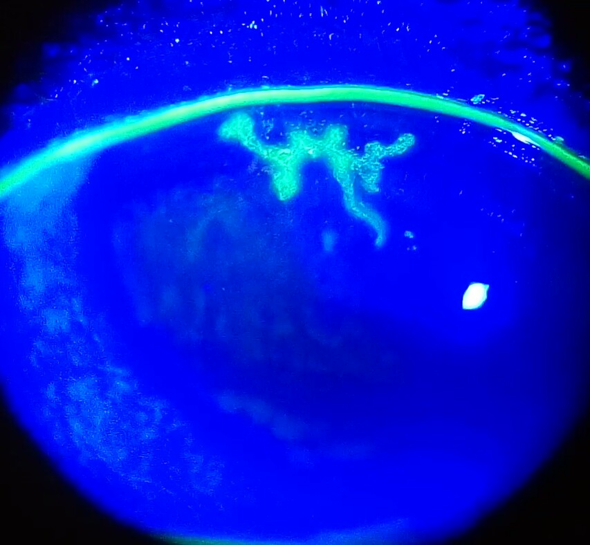
- A branching, tree-like dendritic ulcer with characteristic terminal bulbs, best seen with fluorescein staining and cobalt blue light
- Reduced corneal sensation on gentle testing, a helpful and often overlooked bedside clue
- Stromal haze, infiltrate, or edema in stromal disease, sometimes with an intact overlying epithelium
- Keratic precipitates and corneal edema localized to the endothelium in endotheliitis
- Corneal scarring, thinning, or neovascularization as evidence of prior episodes
A dendritic ulcer with reduced corneal sensation is close to pathognomonic — few other conditions combine both findings.
Diagnostic Evaluation
Clinical Examination
- Diagnosis is primarily clinical, based on the characteristic dendritic pattern on fluorescein staining
- Corneal sensation testing supports the diagnosis and helps distinguish HSV from other causes of epithelial disease
Laboratory Confirmation
- PCR testing of corneal swabs can confirm HSV in atypical or diagnostically uncertain cases
- Viral culture is an alternative but slower confirmatory option, now less commonly used than PCR
Classifying the Disease Form
- Careful slit-lamp assessment to distinguish epithelial, stromal, and endothelial involvement, since this classification directly determines treatment
- Assessment of anterior chamber activity to identify associated uveitis
The diagnostic workup exists mainly to answer one practical question: is this active epithelial infection, or immune-mediated stromal disease?
Differential Diagnosis
Conditions that can be confused with herpes simplex keratitis include:
- Herpes zoster ophthalmicus — pseudodendrites rather than true dendrites, usually with a preceding dermatomal skin rash
- Acanthamoeba keratitis — often associated with contact lens wear and disproportionate pain relative to clinical findings
- Bacterial keratitis — typically presents with a more discrete infiltrate and hypopyon rather than a branching dendritic pattern
- Recurrent corneal erosion — epithelial irregularity related to a prior abrasion rather than a true dendritic ulcer
- Toxic or neurotrophic keratopathy — can mimic epithelial irregularity without the classic dendritic branching pattern
The branching pattern with terminal bulbs, combined with reduced corneal sensation, is usually sufficient to distinguish HSV from these mimics.
Management
Epithelial Keratitis
- Topical or oral antiviral therapy is the mainstay of treatment
- Topical corticosteroids are contraindicated in isolated epithelial disease, since they can enhance viral replication and worsen the ulcer
- Debridement of infected epithelium can be used as an adjunct in select cases
Stromal Keratitis
- Topical corticosteroids, established as beneficial by the Herpetic Eye Disease Study, are used to control the immune-mediated inflammation
- Concurrent antiviral prophylaxis is given alongside steroids to prevent epithelial reactivation while the steroid is used
- Steroid tapering must be gradual, since rapid discontinuation can trigger rebound inflammation
Long-Term Prophylaxis
- Long-term oral antiviral prophylaxis reduces the rate of recurrent epithelial and stromal disease in patients with frequent or severe episodes
- Prophylaxis is particularly appropriate after a first episode of stromal keratitis, given its higher risk of recurrence and scarring
The single most dangerous management error in this disease is using topical corticosteroids on active epithelial dendritic ulceration.
Prognosis
Outcomes depend heavily on which corneal layer is affected and how many prior episodes have occurred.
- Isolated epithelial disease treated promptly with antivirals generally resolves without significant scarring
- Recurrent stromal disease carries a real risk of progressive corneal scarring, neovascularization, and eventual need for corneal transplantation in severe cases
- Reduced corneal sensation from repeated episodes increases the risk of neurotrophic complications independent of active infection
Long-term visual outcome in this disease is driven far more by cumulative recurrences than by any single episode, which is exactly why prophylaxis matters so much after a first significant attack.
Would you like to document anterior segment findings with your smartphone?


Document what you see
Two smartphone imaging tools built for everyday clinic use — one for the slit lamp, one for the fundus.
From Choroida — the team behind this siteReferences
- Herpetic Eye Disease Study Group. A controlled trial of oral acyclovir for the prevention of stromal keratitis or iritis in patients with herpes simplex virus epithelial keratitis. Archives of Ophthalmology. 1997.
- Herpetic Eye Disease Study Group. Oral acyclovir for herpes simplex virus eye disease: effect on prevention of epithelial keratitis and stromal keratitis. Archives of Ophthalmology. 2000.
- American Academy of Ophthalmology. Herpes Simplex Virus Keratitis: A Treatment Guideline. 2014.
- Herpes Simplex Virus Stromal Keratitis and Endotheliitis. EyeWiki, American Academy of Ophthalmology.
- Farooq AV, Shukla D. Herpes simplex epithelial and stromal keratitis: an epidemiologic update. Survey of Ophthalmology. 2012.
Test yourself
A few questions straight from this article.
-
Herpes simplex keratitis is most commonly caused by which virus?
Most ocular herpes simplex disease is caused by HSV-1; HSV-2 occasionally causes ocular disease, particularly in neonates. -
Which form of herpes simplex keratitis represents active viral replication in the cornea?
Epithelial keratitis is active viral replication in the corneal epithelium and produces the classic dendritic ulcer; stromal disease is largely an immune reaction to viral antigen. -
In the Herpetic Eye Disease Study cohort, epithelial keratitis accounted for roughly what share of ocular HSV presentations?
In that landmark cohort epithelial keratitis made up roughly 47% of ocular HSV presentations, while stromal keratitis accounted for roughly 16%. -
Where does herpes simplex virus remain latent between episodes of ocular disease?
After primary infection the virus establishes lifelong latency in the trigeminal ganglion, and on reactivation travels back down the corneal nerves. -
Which slit-lamp finding is characteristic of herpes simplex epithelial keratitis?
The classic sign is a branching, tree-like dendritic ulcer with terminal bulbs, best seen with fluorescein staining under cobalt blue light. -
Which corneal sign supports herpes simplex keratitis but is often overlooked at the bedside?
Reduced corneal sensation on gentle testing is a helpful clue; combined with a dendritic ulcer it is close to pathognomonic, and it can also blunt symptoms and delay presentation. -
Which feature distinguishes herpes zoster ophthalmicus from herpes simplex keratitis?
Herpes zoster ophthalmicus produces pseudodendrites rather than true dendrites, usually preceded by a dermatomal skin rash. -
What is the mainstay of treatment for herpes simplex epithelial keratitis?
Antiviral therapy, topical or oral, is the mainstay for epithelial disease; debridement of infected epithelium can be used as an adjunct in selected cases. -
Why are topical corticosteroids contraindicated in isolated HSV epithelial keratitis?
In active epithelial disease steroids enhance viral replication and worsen the ulcer, which makes this the single most dangerous management error in herpes simplex keratitis. -
How are topical corticosteroids used safely in herpes simplex stromal keratitis?
Steroids control immune-mediated stromal inflammation but require concurrent antiviral prophylaxis to prevent epithelial reactivation, and a gradual taper because abrupt withdrawal can trigger rebound inflammation.

Writes and reviews the clinical articles on Educate. Every post is checked against peer-reviewed sources before it goes live.


