
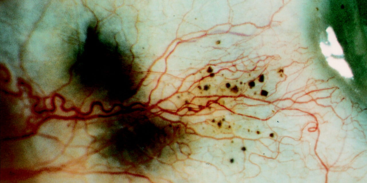
Alkaptonuria is a rare autosomal recessive disorder of tyrosine metabolism, caused by a deficiency of homogentisate 1,2-dioxygenase, leading to accumulation of homogentisic acid and its oxidized polymer, a dark pigment that deposits progressively in connective tissue throughout the body, a process called ochronosis.
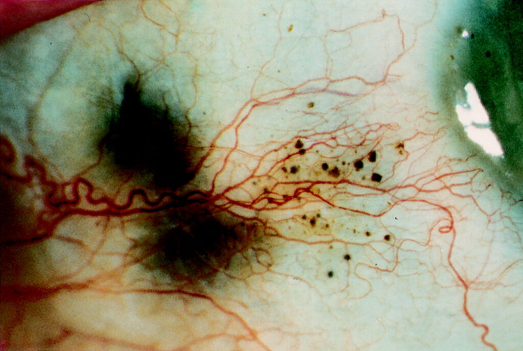
The eye is one of the more visible sites of this pigment deposition, making it a useful, non-invasive clinical window onto a disease process otherwise occurring deep within joints and other internal connective tissue.
Scleral pigmentation is frequently one of the earliest external, easily noticed signs of the underlying systemic disease, sometimes preceding the joint disease that eventually dominates the clinical picture in adulthood.
Because the ocular finding is visible and painless, it is often what a family or clinician first notices, well before the more disabling joint manifestations have had time to develop, giving the eye a genuine early-warning role in a disease that otherwise progresses silently for years.
Pathophysiology
Without functional homogentisate 1,2-dioxygenase, homogentisic acid accumulates in the blood and is excreted in urine, where it oxidizes and polymerizes into a dark, melanin-like pigment on exposure to air.
The classic sign this produces is urine that darkens progressively on standing, sometimes noticed by parents changing an affected infant’s diaper long before any other manifestation of the disease appears.
Over years to decades, the same pigment progressively deposits within collagen-rich connective tissues throughout the body, including cartilage, tendons, skin, and the sclera, producing the characteristic dark discoloration collectively termed ochronosis.
This slow, cumulative deposition process is why the disease is essentially silent in infancy and early childhood beyond the darkening urine, only becoming clinically obvious as visible pigment change and, eventually, joint disease over subsequent decades of life.
Ocular Findings
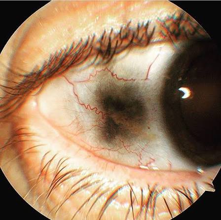
- Scleral pigmentation — brown-black or slate-gray patches, typically most visible in the interpalpebral zone near the horizontal rectus muscle insertions, where the sclera is more exposed and connective tissue is more accessible to the circulating pigment precursor
- Corneal pigmentation, less common than scleral involvement but reported in some patients, typically peripheral and associated with more longstanding, advanced disease
- Conjunctival pigmentation, sometimes accompanying the scleral changes
- The ocular pigmentation itself is generally asymptomatic and does not typically threaten vision, functioning primarily as a visible marker of the underlying systemic disease rather than a source of ocular morbidity in its own right
All-fit Slit-Lamp Adapter
Record and share exactly what you see at the slit lamp. One adapter fits any slit lamp or surgical microscope — and any smartphone.
From Choroida — the team behind this siteSystemic Findings
- Ochronotic arthropathy — a severe, progressive degenerative arthritis affecting the spine and large joints, generally the most functionally significant and disabling manifestation of the disease, developing over decades as pigment accumulates in cartilage and drives progressive joint damage
- Dark pigmentation of the ears (particularly the cartilaginous portions) and, sometimes, other skin areas
- Cardiovascular involvement, including aortic and mitral valve calcification and disease, related to pigment deposition in cardiac connective tissue
- Renal and prostatic stones, related to homogentisic acid and its metabolites
- Darkening of urine on standing — often the earliest, most easily recognized sign, sometimes noted in infancy
Differential Diagnosis of Scleral Pigmentation
- Argyria — diffuse blue-gray pigmentation from chronic silver exposure, discussed in its own dedicated article on this site, with a different pattern and a distinct exposure history rather than an inherited metabolic cause
- Racial/ethnic scleral pigmentation — a normal anatomic variant, generally more uniform and present from birth or early life, without the progressive, patchy, interpalpebral-predominant pattern and without the associated systemic findings of alkaptonuria
- Adrenal insufficiency (Addison’s disease) — can cause mucocutaneous and, less commonly, ocular pigmentary changes, but through an entirely different mechanism (excess ACTH-related melanocyte stimulation) and with its own distinct systemic features (fatigue, hypotension, hyperpigmentation of skin creases and mucous membranes)
- Exogenous pigment deposition from other causes (certain medications, occupational exposures) — distinguished by relevant exposure history
Diagnostic Evaluation
Elevated urinary homogentisic acid confirms the diagnosis, and the classic finding of urine darkening on standing (or more rapidly with alkalinization) can be demonstrated at the bedside as a simple, low-cost supportive test.
Genetic testing for HGD mutations is available and can confirm the diagnosis definitively, useful for family counseling given the autosomal recessive inheritance pattern, particularly for parents considering future pregnancies.
Management
There is no treatment that reverses established ochronotic pigment deposition once it has occurred.
Management is largely supportive and focused on the systemic manifestations, particularly the joint disease, which is managed with standard approaches to degenerative arthritis (pain management, physical therapy, and, in advanced cases, joint replacement surgery) as it progresses over the patient’s lifetime.
Nitisinone, a medication that reduces homogentisic acid production by inhibiting an upstream enzyme in the tyrosine degradation pathway, has shown promise in reducing biochemical markers of disease activity and slowing progression in some studies, representing a disease-modifying option that has changed the management landscape for this previously purely supportive-care condition.
The ocular pigmentation itself requires no specific treatment, since it does not threaten vision, but recognizing it can prompt the broader systemic workup that identifies the underlying diagnosis, in a patient not previously known to have alkaptonuria who presents with otherwise unexplained scleral discoloration.
Once the diagnosis is confirmed, long-term coordinated care with rheumatology, and cardiology given the reported valvular association, becomes the central focus of ongoing management, with the ophthalmologist’s role largely limited to periodic monitoring of the stable, non-progressive ocular finding.


Document what you see
Two smartphone imaging tools built for everyday clinic use — one for the slit lamp, one for the fundus.
From Choroida — the team behind this siteReferences
- Phornphutkul C, Introne WJ, Perry MB, et al. Natural history of alkaptonuria. New England Journal of Medicine.
- Ranganath LR, Jarvis JC, Gallagher JA. Recent advances in management of alkaptonuria. Journal of Clinical Pathology.
- American Academy of Ophthalmology. Basic and Clinical Science Course, Section 8: External Disease and Cornea.
Test yourself
A few questions straight from this article.
-
Deficiency of which enzyme causes alkaptonuria?
Alkaptonuria is a disorder of tyrosine metabolism caused by deficient homogentisate 1,2-dioxygenase, which allows homogentisic acid to accumulate. -
Which inheritance pattern should guide family counseling in alkaptonuria?
Alkaptonuria is autosomal recessive, and HGD genetic testing is particularly useful for parents considering future pregnancies. -
Why does urine in alkaptonuria darken progressively after it is passed?
Excreted homogentisic acid oxidises and polymerises into a dark melanin-like pigment once exposed to air, and alkalinisation speeds the change. -
Where is scleral pigmentation in alkaptonuria typically most visible?
Brown-black or slate-gray patches appear where the sclera is most exposed, typically interpalpebrally near the horizontal rectus muscle insertions. -
A patient has unexplained brown-black scleral patches. What is the chief value of recognising this sign?
The ocular pigment needs no treatment itself, but spotting it in a patient not known to have alkaptonuria triggers the systemic evaluation that makes the diagnosis. -
Which systemic manifestation of alkaptonuria is generally the most functionally disabling?
Pigment accumulating in cartilage over decades drives a severe progressive degenerative arthritis of the spine and large joints, the most disabling feature of the disease. -
Which feature points to argyria rather than alkaptonuria as the cause of scleral pigmentation?
Argyria produces diffuse blue-gray pigmentation and rests on a history of chronic silver exposure rather than an inherited metabolic defect. -
Which cardiovascular complication is associated with alkaptonuria?
Pigment deposition in cardiac connective tissue is linked to aortic and mitral valve calcification and disease, which is why cardiology is part of long-term care. -
Which laboratory finding confirms a diagnosis of alkaptonuria?
Elevated urinary homogentisic acid confirms the diagnosis; HGD genetic testing can confirm it definitively. -
How does nitisinone act as a disease-modifying treatment in alkaptonuria?
Nitisinone inhibits an enzyme upstream in the tyrosine degradation pathway, cutting homogentisic acid production and slowing progression. No treatment reverses pigment already deposited.
