
Interferon retinopathy is a well-recognized, generally reversible retinal microvascular toxicity associated with systemic interferon therapy, most classically encountered historically in patients treated for chronic hepatitis C, though it can occur with interferon used for other indications as well.
Its practical clinical significance lies mainly in recognizing it for what it is — a usually benign, self-limited finding — so that patients are neither unnecessarily frightened nor, conversely, undertreated in the uncommon cases where it does threaten vision, a balance that requires genuine clinical judgment rather than a one-size-fits-all response to every abnormal fundus finding.
Mechanism
Interferon is thought to cause retinal microvascular injury through immune complex deposition and direct microvascular toxicity affecting the retinal capillary network, producing the characteristic combination of small hemorrhages and areas of focal nerve fiber layer ischemia seen on fundus exam.
The precise mechanism remains incompletely understood, but the clinical correlation between interferon dosing and the appearance (and, in most cases, resolution after dose reduction or discontinuation) of the retinal findings supports a direct, dose-related toxic effect on the retinal microvasculature.
Clinical Presentation
The great majority of patients with interferon retinopathy are entirely asymptomatic, with the retinal findings discovered incidentally on a screening or routine dilated fundus exam performed specifically because the patient is on interferon therapy — this asymptomatic pattern is the norm rather than the exception, and is an important point to communicate to anxious patients once the finding is identified.
A small minority of patients, generally those with more extensive retinal involvement or additional risk factors (discussed below), experience visual symptoms related to macular involvement or, rarely, more significant ischemic complications requiring a different, more urgent management approach.
Fundus Explorer Pro
Photograph the retinal findings described here with the phone already in your pocket — 22 D optics and built-in illumination in one handheld unit.
From Choroida — the team behind this siteExam Findings
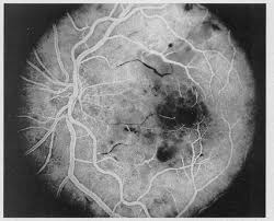
- Cotton wool spots — focal areas of nerve fiber layer infarction, the same basic finding seen in several other retinal vascular conditions discussed elsewhere on this site, here specifically related to interferon-induced microvascular injury
- Retinal hemorrhages, typically flame-shaped or dot-blot in configuration, scattered in the posterior pole
- Findings are usually bilateral, though can be asymmetric, and generally develop within the first few months of starting interferon therapy
- In more severe cases, retinal vascular occlusion, optic disc edema, or macular ischemia can occur, though these more serious complications are distinctly uncommon relative to the milder, typical presentation of scattered cotton wool spots and hemorrhages
Risk Factors for More Severe Disease
- Pre-existing diabetes mellitus — diabetic patients appear to be at higher risk for more significant interferon-related retinal complications, likely reflecting an underlying retinal microvasculature that is already compromised and therefore less able to tolerate an additional toxic insult
- Pre-existing hypertension, for similar reasons related to baseline vascular vulnerability
- Higher interferon dosing and combination therapy regimens
Given this risk pattern, baseline and periodic dilated fundus examination is generally recommended for patients starting interferon therapy who have diabetes or hypertension, allowing early detection of retinopathy before it progresses to a more clinically significant stage, while routine screening in patients without these risk factors is handled somewhat more variably depending on the treating specialists’ practice and the specific clinical context.
Communicating this screening plan clearly to the patient at the start of interferon therapy helps set expectations, since an asymptomatic patient who is asked to attend a dilated eye exam without any visual complaint may otherwise wonder why the visit is necessary.
Differential Diagnosis
- Diabetic retinopathy — can produce a similar picture of cotton wool spots and hemorrhages, and indeed frequently coexists in the same patient given the risk factor overlap; distinguishing purely interferon-related change from underlying diabetic retinopathy can be genuinely difficult and relies partly on the temporal relationship to starting interferon and the pattern of resolution (or lack thereof) with dose adjustment
- Hypertensive retinopathy — a similar basic fundus appearance from a different underlying vascular insult, discussed in this site’s general coverage of that condition, and, again, frequently coexisting given shared risk factors in this patient population
- HIV retinopathy — a similar cotton-wool-spot-predominant picture in a different clinical context (HIV infection rather than interferon exposure), important to distinguish given the different underlying disease and management implications
Management
Most cases of interferon retinopathy require no specific ocular treatment and resolve spontaneously over weeks to a few months, either while interferon therapy continues at the same dose or, in patients with more extensive findings, after dose reduction.
Close monitoring — coordinated between ophthalmology and the treating hepatology, oncology, or infectious disease specialist managing the interferon therapy — allows a shared decision about whether interferon dose adjustment or discontinuation is warranted, weighing the retinal findings against the importance of the underlying systemic treatment being provided, since interrupting an effective systemic therapy is itself a decision with real consequences that should not be made in isolation by ophthalmology alone.
In the uncommon cases with more significant, vision-threatening complications (macular ischemia, disc edema, or retinal vascular occlusion), more urgent discontinuation of interferon therapy is generally warranted, and management of the specific complication follows the same general principles applied to that finding from other causes, discussed in this site’s coverage of retinal vascular occlusion and related ischemic retinal conditions.


Document what you see
Two smartphone imaging tools built for everyday clinic use — one for the slit lamp, one for the fundus.
From Choroida — the team behind this siteReferences
- Hayasaka S, Fujii M, Yamamoto Y, et al. Retinopathy and subconjunctival haemorrhage in patients with chronic viral hepatitis receiving interferon alfa. British Journal of Ophthalmology.
- Schulman JA, Liang C, Kooragayala LM, King J. Posterior segment complications in patients with hepatitis C treated with interferon and ribavirin. Ophthalmology.
- American Academy of Ophthalmology. Basic and Clinical Science Course, Section 12: Retina and Vitreous.
