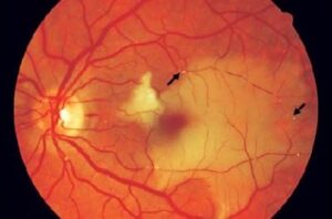
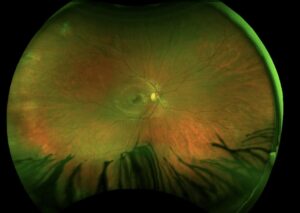
A skin rash on the forehead can tell you exactly what is about to happen inside the eye — if you know where to look.
Herpes zoster ophthalmicus (HZO) is shingles involving the ophthalmic division of the trigeminal nerve, and it is far more than a dermatologic problem.
Roughly one in ten to one in five patients with herpes zoster develop this ophthalmic form.
The eye findings can range from mild conjunctivitis to sight-threatening keratitis, uveitis, and secondary glaucoma.
One physical sign on the tip of the nose can predict much of this risk before the eye is even examined.
Recognizing HZO early, starting antivirals promptly, and knowing which ocular complications to actively screen for is central to preventing permanent vision loss.
What Is Herpes Zoster Ophthalmicus?
Herpes zoster ophthalmicus (HZO) is reactivation of latent varicella-zoster virus within the ophthalmic (V1) division of the trigeminal nerve.
Its defining features include:
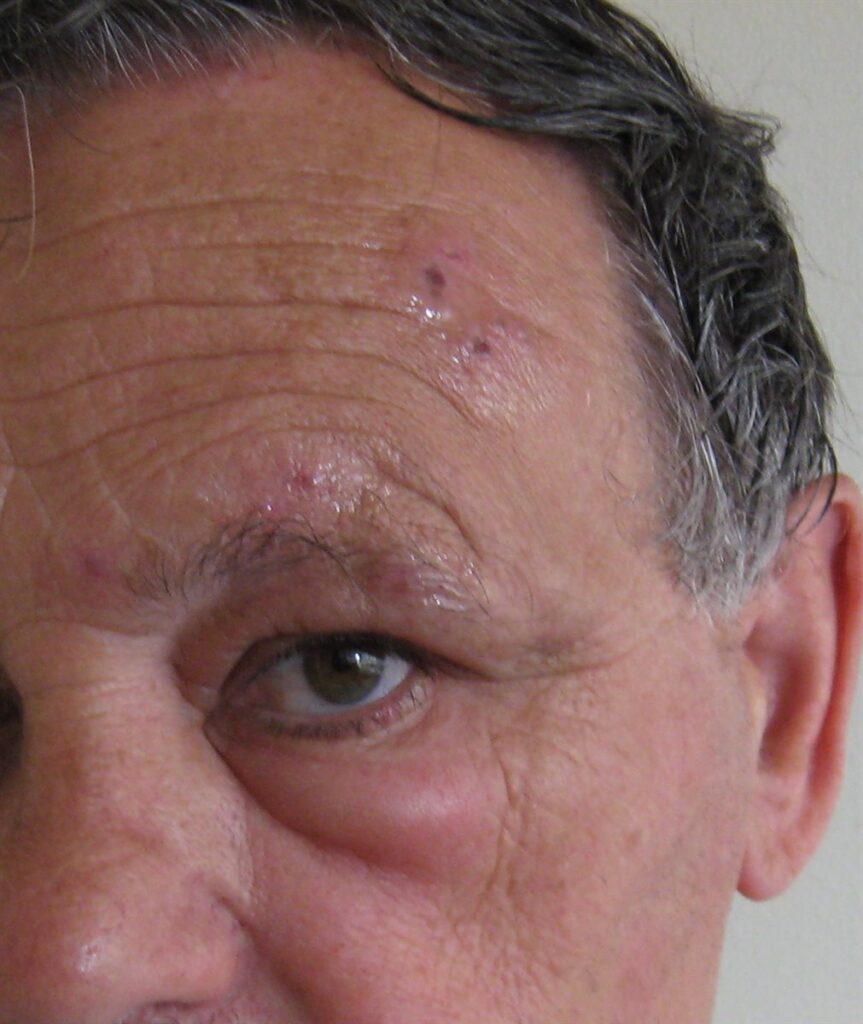
- A painful, vesicular, crusting rash confined to a single dermatome — the forehead, upper eyelid, and scalp on one side
- Sharp respect for the facial midline, since the rash does not cross to the opposite side
- Potential involvement of any ocular structure, from the conjunctiva to the optic nerve
- Development in approximately 4-20% of all patients who develop herpes zoster
Ocular involvement can occur even when the skin rash appears mild, which is why every case of HZO warrants a full eye examination.
Epidemiology
HZO is a recognized and clinically important subset of herpes zoster overall.
- Herpes zoster itself becomes markedly more common with age and with any cause of immunosuppression
- Roughly 4-20% of herpes zoster cases involve the ophthalmic division specifically
- A lesion at the tip of the nose (Hutchinson sign) confers roughly a threefold higher risk of ocular involvement
- Up to 30% of patients who go on to develop ocular involvement do so without a Hutchinson sign, so its absence does not rule out eye disease
Widespread varicella and zoster vaccination has changed the epidemiology of the disease over time, but HZO remains a regular presentation in both primary care and ophthalmology.
All-fit Slit-Lamp Adapter
Record and share exactly what you see at the slit lamp. One adapter fits any slit lamp or surgical microscope — and any smartphone.
From Choroida — the team behind this sitePathophysiology
HZO results from reactivation of virus that has remained dormant since a prior varicella (chickenpox) infection.
- Varicella-zoster virus establishes latency in the trigeminal ganglion after primary infection
- Reactivation, often triggered by waning cell-mediated immunity with age or immunosuppression, allows the virus to travel along the ophthalmic branch of the trigeminal nerve
- Viral replication in the nerve and surrounding tissue produces the characteristic dermatomal skin rash
- The nasociliary nerve, a branch supplying both the tip of the nose and the globe, explains why nasal tip involvement (Hutchinson sign) predicts ocular disease
- Direct viral spread and secondary immune-mediated inflammation can affect any ocular structure along the nerve’s distribution
The shared nerve pathway between the nose and the eye is the anatomical basis for using a skin sign to predict internal ocular risk.
Risk Factors
Patient-Related Risk Factors
- Increasing age, particularly beyond 50-60 years
- Immunosuppression from any cause, including HIV infection, malignancy, and immunosuppressive medication
- Significant physical or psychological stress preceding onset in some patients
- Lack of prior varicella or zoster vaccination
Predictors of Ocular Involvement
- Hutchinson sign — a vesicle or lesion at the tip, side, or root of the nose
- More extensive or severe dermatomal rash
- Delayed initiation of antiviral therapy
Hutchinson sign is a useful red flag, but a completely normal nose does not exclude significant ocular disease.
Clinical Presentation
Symptoms
- Prodromal pain, tingling, or burning in the affected dermatome, often preceding the rash by one to several days
- A painful vesicular rash that progresses to pustules and then crusts over one to two weeks
- Eye pain, redness, photophobia, or blurred vision when ocular structures are involved
- Postherpetic neuralgia, which can persist for months after the skin lesions resolve
Examination Findings
- Vesicular, then crusting, papular skin lesions strictly confined to the V1 dermatome, sparing the opposite side of the face
- Periorbital and eyelid edema, sometimes severe enough to close the eye
- Conjunctival injection, episcleritis, or scleritis in eyes with ocular involvement
- Pseudodendrites on fluorescein staining, distinguishable from the true dendrites of herpes simplex keratitis
- Anterior chamber inflammation, elevated intraocular pressure, or, in severe cases, findings of retinitis or optic neuropathy
Any patient with a V1-distribution zoster rash needs a full slit-lamp exam, including fluorescein staining and IOP check, regardless of how mild the visible eye findings first appear.
Diagnostic Evaluation
Clinical Examination
- Diagnosis is primarily clinical, based on the characteristic dermatomal rash
- Complete ophthalmic exam including visual acuity, IOP, and dilated fundus exam in every patient with V1 zoster
Slit-Lamp and Ocular Surface Assessment
- Fluorescein staining to identify pseudodendrites and assess corneal epithelial involvement
- Assessment for anterior chamber cells and flare indicating anterior uveitis
- Corneal sensation testing, since neurotrophic changes are common and can complicate healing
Laboratory Testing
- Usually unnecessary, since the diagnosis is clinical in typical presentations
- PCR of vesicular fluid can confirm the diagnosis in atypical or diagnostically uncertain cases
The eye exam, not the skin rash alone, is what determines whether a patient needs close ophthalmic follow-up.
Differential Diagnosis
Conditions that can be confused with HZO include:
- Herpes simplex keratitis — true dendrites with terminal bulbs, typically without a dermatomal skin rash
- Contact dermatitis or cellulitis — lacks the characteristic dermatomal, midline-respecting distribution
- Bacterial or viral conjunctivitis — without the preceding dermatomal rash or pseudodendrites
- Preseptal or orbital cellulitis — significant proptosis or restricted motility suggests orbital rather than herpetic disease
The strict dermatomal distribution and midline sparing of the skin rash is usually sufficient to make the diagnosis without additional testing.
Management
Antiviral Therapy
- Oral antivirals — valacyclovir or famciclovir preferred over acyclovir for their more convenient dosing and faster pain resolution — started within 72 hours of rash onset
- Antiviral therapy still provides benefit when started somewhat later, particularly if new lesions are still forming or ocular involvement is present
- Intravenous acyclovir is reserved for severe, disseminated, or immunocompromised presentations
Ocular-Directed Therapy
- Topical corticosteroids for stromal keratitis, episcleritis, scleritis, or anterior uveitis, generally used alongside continued antiviral coverage
- Cycloplegic agents to reduce pain and prevent posterior synechiae in eyes with significant uveitis
- Intraocular pressure-lowering therapy when secondary glaucoma develops from trabeculitis
- Lubrication and, when needed, specific neurotrophic keratitis management for eyes with reduced corneal sensation
Long-Term and Preventive Measures
- Zoster vaccination is recommended for eligible adults to reduce the incidence and severity of herpes zoster, including HZO
- Longer-term, low-dose suppressive valacyclovir has been shown in recent trial data to reduce disease flares and new or worsening ocular complications over the following months
- Management of postherpetic neuralgia, which can be more disabling than the acute episode itself
Prompt antiviral treatment combined with a genuinely complete eye exam is what prevents the majority of HZO’s most serious ocular complications.
Prognosis
Outcomes depend heavily on how early treatment starts and which ocular structures become involved.
- Most patients treated promptly with antivirals have a favorable outcome, with the skin rash resolving over several weeks
- Chronic or recurrent keratitis, uveitis, and secondary glaucoma can develop months to years after the acute episode, sometimes requiring long-term follow-up
- Postherpetic neuralgia can persist well beyond resolution of the rash and significantly affect quality of life
HZO is not a disease that ends when the rash clears — ongoing risk of recurrent ocular inflammation makes long-term follow-up appropriate even after the acute episode resolves.
Would you like to document anterior segment findings with your smartphone?


Document what you see
Two smartphone imaging tools built for everyday clinic use — one for the slit lamp, one for the fundus.
From Choroida — the team behind this siteReferences
- Zoster Eye Disease Study (ZEDS) Research Group. Effect of long-term suppressive valacyclovir on herpes zoster ophthalmicus. JAMA Ophthalmology. 2024.
- Herpes Zoster Ophthalmicus. StatPearls, NCBI Bookshelf.
- Tran KD, Falcone MM, Choi DS, et al. Epidemiology of herpes zoster ophthalmicus: recurrence and chronicity. Ophthalmology. 2016.
- Vrcek I, Choudhury E, Durairaj V. Herpes zoster ophthalmicus: a review for the internist. American Journal of Medicine. 2017.
- Herpes Zoster Ophthalmicus. EyeWiki, American Academy of Ophthalmology.
Test yourself
A few questions straight from this article.
-
Herpes zoster ophthalmicus results from viral reactivation in which nerve division?
HZO is reactivation of latent varicella-zoster virus that has lain dormant in the trigeminal ganglion, travelling along the ophthalmic (V1) division to produce the dermatomal rash. -
What proportion of herpes zoster cases involve the ophthalmic division of the trigeminal nerve?
Roughly 4-20% of all herpes zoster patients develop the ophthalmic form, which is why HZO remains a regular presentation in both primary care and ophthalmology. -
What is the Hutchinson sign in herpes zoster ophthalmicus?
A vesicle or lesion on the tip, side or root of the nose is the Hutchinson sign, and it confers roughly a threefold higher risk of ocular involvement. -
Which nerve explains why nasal tip lesions predict ocular disease in herpes zoster ophthalmicus?
The nasociliary nerve supplies both the tip of the nose and the globe, so involvement of that shared branch links a skin sign to internal ocular risk. -
What proportion of patients with ocular involvement from zoster have no Hutchinson sign?
Up to 30% of patients who develop ocular involvement do so without any Hutchinson sign, so a normal nose never excludes eye disease. -
Which corneal finding on fluorescein staining suggests zoster rather than herpes simplex keratitis?
Zoster produces pseudodendrites, which are distinguishable from the true dendrites with terminal bulbs seen in herpes simplex keratitis. -
Within what window should oral antivirals ideally begin in herpes zoster ophthalmicus?
Oral valacyclovir or famciclovir should start within 72 hours of rash onset, though later treatment still helps if new lesions are forming or the eye is involved. -
Which herpes zoster ophthalmicus presentation warrants intravenous acyclovir?
Intravenous acyclovir is reserved for severe, disseminated or immunocompromised presentations; most other patients are managed with oral antivirals. -
Secondary glaucoma in herpes zoster ophthalmicus develops through which mechanism?
Inflammation of the trabecular meshwork raises intraocular pressure in HZO, and pressure-lowering therapy is added when this secondary glaucoma develops. -
Which strategy reduces later flares and new ocular complications after herpes zoster ophthalmicus?
Recent trial data show that longer-term, low-dose suppressive valacyclovir reduces disease flares and new or worsening ocular complications in the months after the acute episode.

Writes and reviews the clinical articles on Educate. Every post is checked against peer-reviewed sources before it goes live.