
An idiopathic full-thickness macular hole is a defect that extends through all layers of the retina at the fovea, and it is one of the few retinal conditions where a well-timed surgery restores meaningful vision in a genuinely predictable way. Most cases arise from anteroposterior traction exerted by the vitreous as it separates from the retina with age, concentrated at the fovea because that is where vitreomacular adhesion tends to be strongest.
The condition is distinct from lamellar macular holes and from epiretinal membrane–related pseudoholes, both of which spare at least some retinal thickness and behave very differently in terms of both natural history and surgical urgency, making the distinction genuinely important to get right before counseling a patient about their prognosis.
Pathogenesis
Gass’s original staging system, later refined with OCT-era understanding, describes progression from an impending hole (a foveal cyst with persistent vitreofoveal traction) through a small full-thickness defect to a large hole with an operculum floating in the vitreous. Not every impending hole progresses — a meaningful proportion resolve spontaneously if the vitreous releases before the traction causes a full-thickness break.
Once the hole is full-thickness, spontaneous closure is uncommon, though it does happen occasionally in small holes, which is part of why size at presentation matters so much for counseling and surgical timing.
Clinical Presentation
Patients describe gradual, painless central visual distortion or blurring, often first noticed when covering the unaffected eye. Metamorphopsia — straight lines appearing bent or wavy — is a common early complaint and can be demonstrated at the bedside with an Amsler grid.
A central or paracentral scotoma develops as the hole enlarges, and visual acuity typically falls into the 20/60 to 20/200 range by the time patients present with a stage 3 or 4 hole, though this varies with hole size and duration.
Fundus Explorer Pro
Photograph the retinal findings described here with the phone already in your pocket — 22 D optics and built-in illumination in one handheld unit.
From Choroida — the team behind this siteExam and Imaging Findings
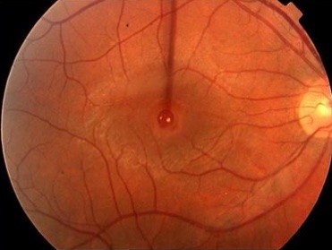
- A round, reddish foveal defect on fundus examination, sometimes with a surrounding cuff of subretinal fluid
- Positive Watzke-Allen sign — a thin slit-beam appears broken or narrowed as it crosses the hole
- Yellow deposits at the base of the hole in some cases
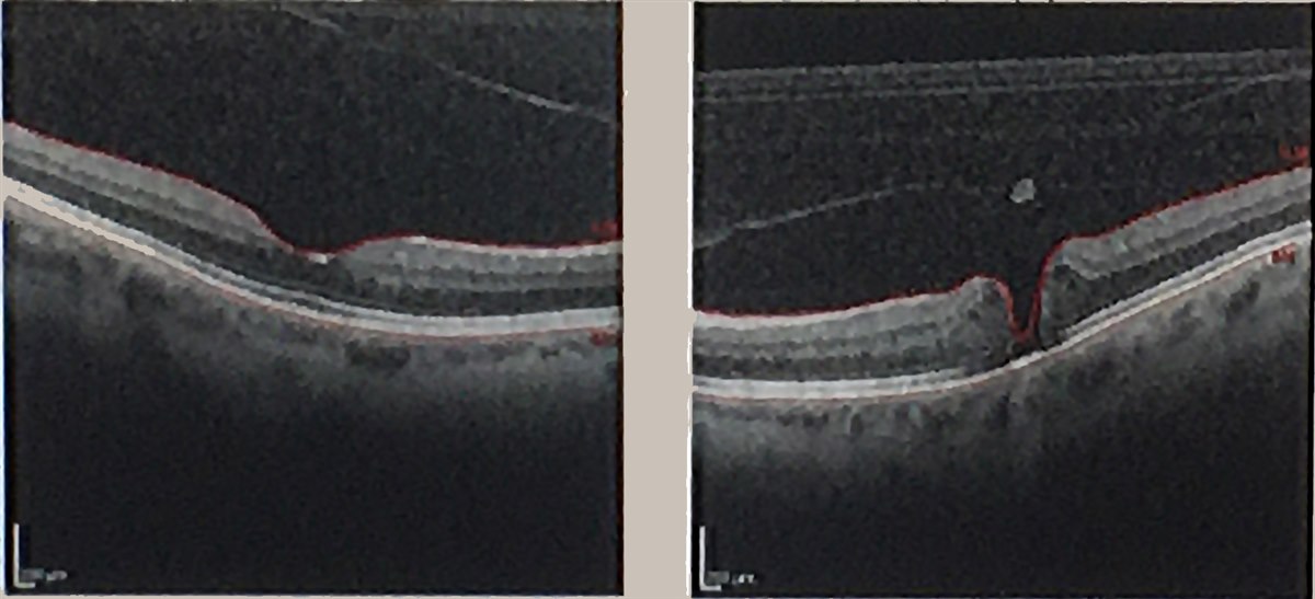
- OCT showing the full-thickness neurosensory defect, which has become the primary tool for staging and measuring hole diameter
OCT has largely replaced clinical staging alone, since it measures the minimum linear diameter of the hole directly — a number that correlates closely with both surgical success rate and the likelihood of good postoperative vision, making it central to both surgical planning and patient counseling.
Differential Diagnosis
- Lamellar macular hole — partial-thickness defect, epiretinal membrane commonly present, generally stable
- Macular pseudohole — steepened foveal contour from epiretinal membrane traction without an actual retinal defect
- Vitreomacular traction — foveal distortion without a full-thickness break
- Solar retinopathy — history of sun gazing, foveal outer retinal defect, no true hole
- Cystoid macular edema — cystic spaces without a full-thickness dehiscence
Management
Pars plana vitrectomy with induction of posterior vitreous detachment, peeling of the internal limiting membrane, and gas tamponade is the standard surgical approach for holes that are unlikely to close spontaneously — essentially all stage 2 holes and larger. Face-down positioning after surgery was traditionally recommended to keep the gas bubble in contact with the macula, though modern data suggest the requirement can often be relaxed, particularly for smaller holes, without meaningfully compromising closure rates.
Ocriplasmin, an intravitreal enzymatic vitreolysis agent, can be used for smaller holes with focal vitreomacular traction and no significant epiretinal membrane, avoiding surgery in carefully selected cases, though its overall closure rates are lower than surgery and its use has narrowed as surgical outcomes have continued to improve. For very small, early holes, observation with serial OCT is reasonable given the real chance of spontaneous resolution.
The choice between these three pathways — observation, pharmacologic vitreolysis, or surgery — depends heavily on hole size, the presence of coexisting vitreomacular traction or epiretinal membrane, and how the specific anatomic features on OCT map onto what each treatment option is actually designed to address.
Prognosis
Anatomic closure rates with modern vitrectomy techniques exceed 90 percent for most holes, and closure rates are inversely related to preoperative hole size — smaller holes close more reliably and recover better vision. Visual recovery lags behind anatomic closure by weeks to months as the photoreceptor layer remodels, and some patients are left with a persistent, if smaller, central scotoma or metamorphopsia even after a successfully closed hole, which is a useful point to set expectations around before surgery rather than only discovering it postoperatively.
Holes that have been present longer before surgery, and holes associated with high myopia or significant epiretinal membrane, tend to have a harder time closing and a more limited visual recovery even when they do close. This is the practical argument for referring suspected macular holes promptly rather than watching them for an extended period, since delay carries a genuine, measurable cost to both anatomic and visual outcomes rather than simply preserving the option of treatment for later.


Document what you see
Two smartphone imaging tools built for everyday clinic use — one for the slit lamp, one for the fundus.
From Choroida — the team behind this siteReferences
- Gass JD. Idiopathic senile macular hole: its early stages and pathogenesis. Archives of Ophthalmology.
- Duker JS, et al. The International Vitreomacular Traction Study Group classification of vitreomacular adhesion, traction, and macular hole. Ophthalmology.
- Kelly NE, Wendel RT. Vitreous surgery for idiopathic macular holes. Archives of Ophthalmology.
- Ip MS, et al. Anatomical outcomes of surgery for idiopathic macular hole as determined by optical coherence tomography. Archives of Ophthalmology.
- Stalmans P, et al. Enzymatic vitreolysis with ocriplasmin for vitreomacular traction and macular holes. New England Journal of Medicine.
Test yourself
A few questions straight from this article.
-
What mechanism produces most idiopathic full-thickness macular holes?
The vitreous exerts this traction as it separates with age, concentrated at the fovea because vitreomacular adhesion is strongest there. -
What characterises an impending, stage 1 macular hole?
A meaningful proportion of these resolve spontaneously if the vitreous releases before traction causes a full-thickness break. -
What defines a positive Watzke-Allen sign in macular hole?
The beam looks broken or narrowed as it crosses the defect, alongside a round reddish foveal lesion sometimes cuffed by subretinal fluid. -
Which OCT measurement best predicts surgical success in full-thickness macular hole?
This directly measured number correlates closely with both surgical success rate and the likelihood of good postoperative vision. -
What visual acuity do patients with a stage 3 or 4 macular hole typically present with?
Acuity varies with hole size and duration, and a central or paracentral scotoma develops as the hole enlarges. -
What defines a macular pseudohole?
The appearance comes from epiretinal membrane traction alone; no retinal tissue is actually missing, unlike a true full-thickness hole. -
Which procedure is the standard approach for a stage 2 or larger macular hole?
Posterior vitreous detachment is induced at the same time; face-down positioning afterwards can often be relaxed, particularly for smaller holes. -
In which situation is ocriplasmin a reasonable alternative to macular hole surgery?
Enzymatic vitreolysis avoids surgery in carefully selected cases, but its closure rates are lower and its use has narrowed as surgery improved. -
What anatomic closure rate does modern vitrectomy achieve for most macular holes?
Closure rates are inversely related to preoperative hole size, and visual recovery lags behind closure by weeks to months as photoreceptors remodel. -
Which factors make a macular hole harder to close with more limited visual recovery?
This is the practical argument for prompt referral, since delay carries a measurable cost to both anatomic and visual outcomes.

Writes and reviews the clinical articles on Educate. Every post is checked against peer-reviewed sources before it goes live.


