
CASE REPORT
A 5-year-old male, presented with facial asymmetry, right-sided microtia (underdeveloped ear), and ocular abnormalities. The ocular findings included eyelid coloboma (a gap or notch in the eyelid), microphthalmia (abnormally small eye), and epi-bulbar dermoid (a benign tumor-like growth on the surface of the eye).


The patient’s medical history revealed no significant ocular or systemic conditions. Ocular examination was conducted, including visual acuity assessment, slit-lamp examination, dilated fundus examination, and ocular motility evaluation.
The visual acuity in the affected eye was reduced compared to the unaffected eye. The slit-lamp examination revealed the presence of an epibulbar dermoid, causing corneal astigmatism and irregularity. The fundus examination did not show any significant abnormalities.
According to the results of the previous examination, the patient was diagnosed with Goldenhar syndrome.
Goldenhar Syndrome DISEASE entity
Goldenhar syndrome, Also called Facio-auricula-vertebral dysplasia, unilateral craniofacial microsomia, first and second branchial arch syndrome, lateral facial dysplasia, otomandibular dysostosis, velocardiofacial syndrome, and unilateral mandibulofacial dysostosis.

Goldenhar syndrome is characterized by craniofacial anomalies in association with vertebral, cardiac, renal, and central nervous system defects. The syndrome is characterized by a triad of anomalies comprising epibulbar dermoid, accessory auricular appendages, and aural fistula.
Various hypothesis has been put forward to explain etiopathogenesis. Gorlin and Pindborg suggested that an unknown primary cause of faulty embryological development causes an abnormality of the mesoblasts giving rise to branchial and vertebral anomalies.
Jong Bloet hypothesized that the syndrome was a result of fertilization of the overripe or aged ovum. Currently accepted theory states that the syndrome occurs due to an imbalance in cells during the blastogenesis period (30-45 days of intrauterine life) of embryo formation.
It is found to involve the derivatives of first and second branchial arches. The condition is apparent at birth, but the phenotype can vary greatly in its severity depending on the activation and expression of the defective gene.
Goldenhar Syndrome MANAGEMENT
The ocular dermoid are benign tumors but may lead to serious ophthalmic consequences like induced astigmatism, amblyopia, and strabismus. The epibulbar lipodermoids and dermoids of large size may obstruct the visual axis, cause limitation of ocular motility or hinder eyelid closure leading to exposure keratitis and corneal ulceration.
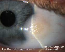
The dermoid with lashes may cause constant rubbing of the eye leading to symptoms of irritation, watering, discharge, secondary infections, and corneal epithelial defects and ulcerations. The early diagnosis and surgical excision of limbal dermoid lesions may prevent amblyopia and strabismus, especially in the pediatric age group.
Eyelid reconstruction of a coloboma may often be challenging due to the small facial structures and limited movement of surrounding soft tissues. Eyelid-sharing techniques for eyelid margin reconstruction are often not ideal due to the risk of occlusion amblyopia.
Surgery
Reconstructive surgery can be performed for structural deformities of the eyes and ears.

Stem cell grafting (womb tissue grafting) has been successfully used to “reprogram” eye dermoids, effectively halting the regrowth of eye dermoids.
HOW TO TAKE SLIT-LAMP EXAM IMAGES WITH A SMARTPHONE?
Smartphone slit-lamp photography is the new advancement in the field of science and technology in which photographs of the desired slit-lamp finding can be taken with smartphones by using the slit-lamp adapters.
Slit-lamp Smartphone photography
REFERENCES
- American Academy of Ophthalmology. Goldenhar-Gorlin syndrome.
- Gorlin RJ, Cohen MM, Hennekam RC. Syndromes of the Head and Neck. 4th ed. New York: Oxford University Press; 2001.
- Kokavec R. Goldenhar syndrome with various clinical manifestations. Cleft Palate Craniofac J 2006;43:628-34.
- Bekibele CO, Ademola SA, Amanor-Boadu SD, Akang EE, Ojemakinde KO. Goldenhar syndrome: A case report and literature review. West Afr J Med 2005;24:77-80.
- Madiyal A, Babu SG, Ajila V, Madi M, Bhat S. Goldenhar syndrome: Report of two cases with review of literature. CHRISMED J Health Res 2018;5:67-71.
- Sinha S, Singh AK, Mehra A, Singh R. Goldenhar syndrome – A literature review. JSM Dent 2015;3:1052-5.
Slit-lamp Smartphone photography

RETINAL IMAGING BY YOUR SMARTPHONE