
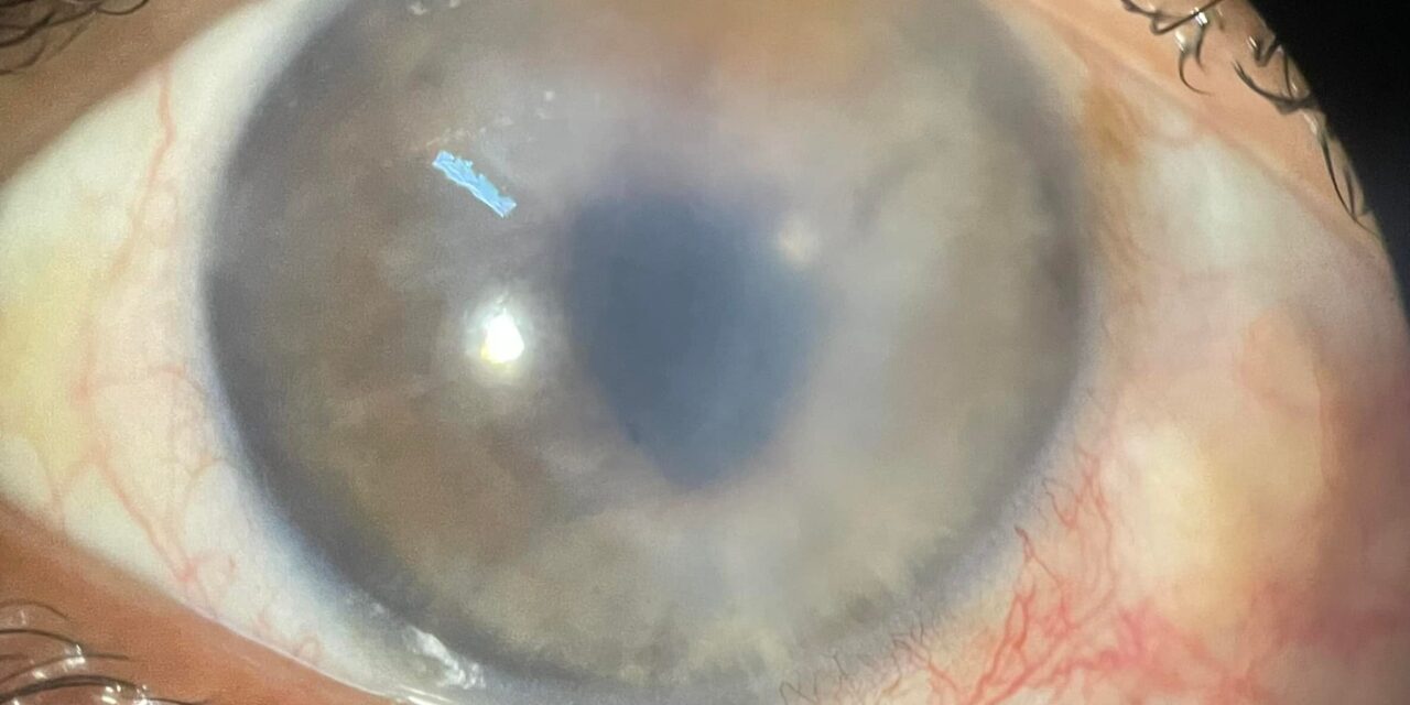
Case Study
A 72-year-old pseudophakic patient presented with progressive blurred vision, ocular discomfort, and recurrent episodes of foreign body sensation several months after uncomplicated cataract surgery.
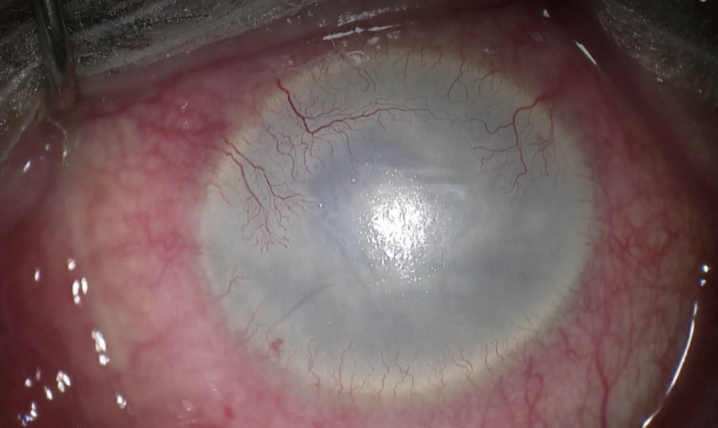
Visual acuity was markedly reduced, and symptoms were worse in the morning. Slit-lamp examination revealed corneal edema with multiple epithelial bullae and Descemet membrane folds.
Specular microscopy demonstrated significantly reduced endothelial cell density. Anterior segment OCT confirmed stromal thickening without evidence of intraocular inflammation or raised intraocular pressure.
Based on the clinical findings and surgical history, a diagnosis of pseudophakic bullous keratopathy was established.
Disease Entity
Pseudophakic bullous keratopathy (PBK) is a form of irreversible corneal edema that occurs due to endothelial cell failure following cataract surgery with intraocular lens implantation.
It represents one of the most common indications for endothelial keratoplasty worldwide.
The condition arises when endothelial cell loss exceeds the cornea’s compensatory capacity, leading to chronic stromal and epithelial edema.
All-fit Slit-Lamp Adapter
Record and share exactly what you see at the slit lamp. One adapter fits any slit lamp or surgical microscope — and any smartphone.
From Choroida — the team behind this sitePathophysiology
The corneal endothelium maintains corneal transparency through active fluid transport from the stroma into the anterior chamber.
Cataract surgery can damage endothelial cells through mechanical trauma, ultrasound energy, irrigation fluids, or postoperative inflammation.
When endothelial cell density falls below a critical threshold, endothelial pump function fails. As a result, stromal hydration increases, collagen lamellae separate, and corneal thickness rises.
Chronic edema causes epithelial bullae formation, which may rupture and result in significant pain.
Etiology and Risk Factors
Several factors increase the risk of developing pseudophakic bullous keratopathy:
-
Pre-existing low endothelial cell count
-
Fuchs endothelial corneal dystrophy
-
Complicated cataract surgery
-
Prolonged phacoemulsification time
-
Excessive ultrasound energy
-
Intraoperative endothelial trauma
-
Intraocular lens malposition or endothelial touch
Older age and repeated intraocular surgeries further raise the risk.
Clinical Features
Patients with PBK typically present with both visual and symptomatic complaints.
Symptoms
-
Progressive blurred vision
-
Glare and halos
-
Ocular pain from ruptured bullae
-
Foreign body sensation
-
Photophobia
-
Excessive tearing
Signs
-
Diffuse corneal edema
-
Epithelial and subepithelial bullae
-
Descemet membrane folds
-
Stromal haze
-
Reduced corneal transparency
Symptoms often fluctuate, with worse vision upon awakening.

Examination Findings
-
Visual acuity: Reduced, often severely
-
Slit-lamp exam: Corneal edema with bullae
-
Specular microscopy: Low endothelial cell density
-
Pachymetry: Increased central corneal thickness
-
Anterior segment OCT: Stromal and epithelial edema
Differential Diagnosis
Several conditions may mimic pseudophakic bullous keratopathy:
-
Fuchs endothelial corneal dystrophy
-
Aphakic bullous keratopathy
-
Chronic uveitis-related corneal edema
-
Herpetic endotheliitis
-
Corneal decompensation from glaucoma
A careful surgical history is essential for diagnosis.
Diagnosis
PBK is primarily a clinical diagnosis based on patient history, slit-lamp findings, and evidence of endothelial dysfunction. Ancillary testing helps confirm severity and guide management, but is not always required.
Management
Management depends on disease severity and symptom burden.
Medical Management
Conservative therapy aims to relieve symptoms but does not reverse endothelial failure.
-
Hypertonic saline drops and ointment
-
Lubricating eye drops
-
Bandage contact lenses for pain control
-
Topical antibiotics if epithelial defects are present
These measures offer temporary relief only.
Surgical Management
Definitive treatment requires corneal transplantation.
-
Descemet stripping endothelial keratoplasty (DSEK)
-
Descemet membrane endothelial keratoplasty (DMEK)
-
Penetrating keratoplasty (in advanced cases)
Endothelial keratoplasty has largely replaced full-thickness grafts due to faster recovery and better visual outcomes.
Prognosis
Visual prognosis depends on timely intervention. Early endothelial keratoplasty can restore good visual acuity. Delayed treatment may result in irreversible stromal scarring and suboptimal outcomes. Pain relief is usually achieved after successful surgery.
Prevention
Preventive strategies focus on minimizing endothelial damage during cataract surgery:
-
Preoperative endothelial assessment
-
Gentle surgical technique
-
Reduced phacoemulsification energy
-
Adequate viscoelastic use
-
Early recognition of endothelial compromise


Document what you see
Two smartphone imaging tools built for everyday clinic use — one for the slit lamp, one for the fundus.
From Choroida — the team behind this siteReferences
-
Bourne WM. Biology of the corneal endothelium in health and disease. Eye.
-
Price MO, Price FW. Endothelial keratoplasty—A review. Clinical & Experimental Ophthalmology.
-
Melles GRJ, Ong TS, Ververs B, van der Wees J. Descemet membrane endothelial keratoplasty (DMEK). Cornea.
-
Lass JH, Benetz BA, Gal RL. Corneal endothelial cell loss after cataract surgery. Ophthalmology.
-
Patel SV. Corneal edema and endothelial dysfunction after intraocular surgery. American Journal of Ophthalmology.
-
Eye Bank Association of America. Indications for corneal transplantation. EBAA Statistical Report.
Test yourself
A few questions straight from this article.
-
What underlies the corneal oedema of pseudophakic bullous keratopathy?
PBK is irreversible corneal oedema that follows endothelial cell failure after cataract surgery with lens implantation, once cell loss outstrips the cornea's compensatory capacity. -
How does the healthy corneal endothelium keep the cornea transparent?
The endothelium pumps fluid out of the stroma and into the anterior chamber; when that pump fails, stromal hydration rises and the cornea clouds. -
Which intraoperative factor raises the risk of pseudophakic bullous keratopathy?
Prolonged phacoemulsification time and excessive ultrasound energy damage endothelial cells during surgery, alongside mechanical trauma, irrigation fluids and postoperative inflammation. -
Vision in pseudophakic bullous keratopathy characteristically follows which daily pattern?
Symptoms fluctuate and are typically worse on waking, when overnight lid closure leaves the oedematous cornea at its thickest. -
Which finding accounts for the ocular pain in pseudophakic bullous keratopathy?
Chronic oedema drives epithelial bullae formation, and it is the rupture of these bullae that produces significant pain and foreign body sensation. -
Which test directly demonstrates the endothelial deficit in pseudophakic bullous keratopathy?
Specular microscopy shows the reduced endothelial cell density that defines the disease, while pachymetry and anterior segment OCT only document the resulting oedema. -
Which feature best separates pseudophakic bullous keratopathy from Fuchs endothelial corneal dystrophy?
The two share oedema, bullae and low cell counts, so a careful surgical history showing preceding cataract surgery is what establishes the diagnosis of PBK. -
What do hypertonic saline drops achieve in pseudophakic bullous keratopathy?
Conservative measures such as hypertonic saline, lubricants and bandage lenses ease symptoms but do nothing to reverse the underlying endothelial failure. -
Why has endothelial keratoplasty largely replaced penetrating keratoplasty for pseudophakic bullous keratopathy?
DSEK and DMEK replace only the failed endothelial layer, giving faster recovery and better visual results than a full-thickness graft. -
Why does timing matter in the surgical management of pseudophakic bullous keratopathy?
Early endothelial keratoplasty can restore good acuity, whereas delay lets irreversible stromal scarring develop and caps the visual result.
