
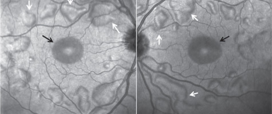
MEK inhibitor-associated retinopathy (MEKAR) is a drug-induced retinopathy that causes multifocal serous subretinal fluid in patients treated with MEK inhibitors for cancer.
It appears within days to weeks of starting therapy, is often bilateral, and usually resolves without permanent visual loss.
Oncologists rely on ophthalmologists to distinguish it from more serious causes of visual disturbance, and to advise whether therapy needs to change.
What Is MEK Inhibitor-Associated Retinopathy?
MEK1 and MEK2 are kinases in the RAS-RAF-MEK-ERK signaling pathway.
MEK inhibitors block this pathway and are used in several cancers.
- Trametinib, cobimetinib, and binimetinib are used with BRAF inhibitors in BRAF-mutant melanoma and other tumors
- Selumetinib is used for plexiform neurofibromas in neurofibromatosis type 1 (see neurofibromatosis type 1)
- Other MEK inhibitors are used in lung cancer, low-grade glioma, and other malignancies
Ocular toxicity is common in trials, and MEKAR is among the most frequent ocular adverse effects.
Pathogenesis
The exact mechanism is not certain.
MEK signaling is important for RPE function and for the maintenance of tight junctions and fluid transport.
Inhibition of this pathway is thought to impair RPE pump function and increase subretinal fluid accumulation.
The pattern differs from central serous chorioretinopathy, where choroidal hyperpermeability plays a major role.
Fundus Explorer Pro
Photograph the retinal findings described here with the phone already in your pocket — 22 D optics and built-in illumination in one handheld unit.
From Choroida — the team behind this siteClinical Features
Patients may report blurred vision, metamorphopsia, photopsia, or a change in color perception, and some are asymptomatic.
Onset is usually within the first weeks of treatment, and in some series the median time was about a month.
Findings include:
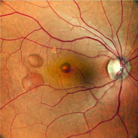
- Bilateral, multifocal, small serous detachments of the neurosensory retina, often involving the fovea
- Shallow subretinal fluid with a normal RPE contour
- Absence of leakage on fluorescein angiography in many cases
- Preserved or mildly reduced visual acuity
OCT shows subfoveal or parafoveal fluid, sometimes with subretinal hyperreflective material and mild outer retinal thickening.
Other reported ocular effects include retinal vein occlusion, uveitis, dry eye, and blepharitis.
Differential Diagnosis
- Central serous chorioretinopathy, which is usually unilateral and shows leakage on angiography and choroidal thickening (see central serous chorioretinopathy)
- Vogt-Koyanagi-Harada disease and other inflammatory serous detachments (see VKH disease)
- Retinal vein occlusion, also reported with MEK inhibitors
- Metastatic disease to the choroid or retina, which needs to be excluded in cancer patients
- Other drug toxicity, including hydroxychloroquine, tamoxifen, and immune checkpoint inhibitor-related uveitis
The presence of bilateral, symmetrical, shallow foveal detachments in a patient on a MEK inhibitor is characteristic.
Management
Coordination With Oncology
Management is a shared decision with the oncologist.
Most cases are mild and do not require stopping treatment.
Observation
When vision is good and symptoms are mild, observation with repeat OCT is reasonable.
The fluid often resolves spontaneously even when the drug is continued.
Dose Interruption or Reduction
If vision drops or the subretinal fluid is extensive or persistent, the drug can be interrupted or the dose reduced.
Resolution typically follows within weeks of interruption, and treatment can often be resumed at a lower dose.
Screening
Baseline ophthalmic examination is recommended before starting therapy, and patients should be told to report visual symptoms promptly.
Repeat examination is advised if symptoms develop, with OCT to detect fluid.
Other Complications
Retinal vein occlusion needs standard management, including treatment of macular edema and a discussion with oncology about continuing the drug.
Uveitis is treated with topical steroids and cycloplegics.
Monitoring and Counseling
Practical points help make the collaboration with oncology work.
- Perform baseline visual acuity, dilated fundus examination, and OCT before or soon after starting a MEK inhibitor
- Ask patients to report blurred vision, distortion, or flashes the same week they occur
- Repeat OCT when symptoms appear, and compare with baseline to identify new fluid
- Send a short written summary to the oncologist stating whether the finding is consistent with MEKAR, whether vision is threatened, and whether a drug change is advised
- Avoid unnecessary steroid treatment, which is not helpful in MEKAR and may cause harm
Patients are reassured to learn that the fluid is a known effect of the drug, that vision usually recovers, and that the cancer treatment can often continue.
They should still be told that new visual symptoms need review, since retinal vein occlusion and other causes of visual loss can occur on the same drugs.
Other Ocular Effects of Targeted Cancer Therapy
Patients on MEK inhibitors, often combined with BRAF inhibitors, may develop other ocular problems.
- Uveitis, more often reported with BRAF inhibitors and with combination therapy
- Retinal vein occlusion, which is uncommon but potentially sight-threatening (see central retinal vein occlusion)
- Dry eye, blepharitis, and conjunctival irritation
- Optic neuropathy, reported occasionally
Immune checkpoint inhibitors produce a different set of toxicities, including uveitis, VKH-like disease, and optic neuritis, so the drug regimen should be known before assigning a cause to a new visual symptom.
Patients on any of these agents benefit from a baseline ophthalmic examination, and they should have quick access to review if symptoms develop.
Prognosis
The retinopathy is usually mild and self-limited.
Visual acuity typically returns to baseline, and permanent visual loss is rare.
Some patients have recurrent fluid when the drug is restarted.
Awareness that this is a known and benign class effect prevents unnecessary investigation, biopsy, or discontinuation of effective cancer therapy.


Document what you see
Two smartphone imaging tools built for everyday clinic use — one for the slit lamp, one for the fundus.
From Choroida — the team behind this siteReferences
- Francis JH, Habib LA, Abramson DH, et al. Clinical and morphologic characteristics of MEK inhibitor-associated retinopathy: differences from central serous chorioretinopathy. Ophthalmology. 2017;124:1788-1798.
- Urner-Bloch U, Urner M, Stieger P, et al. Transient MEK inhibitor-associated retinopathy in metastatic melanoma. Ann Oncol. 2014;25:1437-1441.
- Stjepanovic N, Velazquez-Martin JP, Bedard PL. Ocular toxicities of MEK inhibitors and other targeted therapies. Ann Oncol. 2016;27:998-1005.
- Liu CY, Francis JH, Brodie SE, et al. Retinal toxicities of cancer therapy drugs: biologics, small molecule inhibitors, and chemotherapies. Retina. 2014;34:1261-1280.
- Renouf DJ, Velazquez-Martin JP, Simpson R, Siu LL, Bedard PL. Ocular toxicity of targeted therapies. J Clin Oncol. 2012;30:3277-3286.
Test yourself
A few questions straight from this article.
-
Which retinal change characterises MEK inhibitor-associated retinopathy?
MEKAR produces bilateral, multifocal, small serous detachments of the neurosensory retina, often involving the fovea, with shallow subretinal fluid and a normal RPE contour. -
MEK inhibitors act on which intracellular signalling pathway?
MEK1 and MEK2 are kinases in the RAS-RAF-MEK-ERK signalling pathway, and MEK inhibitors block this pathway in the treatment of several cancers. -
What is thought to cause the subretinal fluid in MEK inhibitor-associated retinopathy?
MEK signalling maintains RPE tight junctions and fluid transport, so blocking the pathway is thought to impair the RPE pump and allow subretinal fluid to accumulate. -
When does MEK inhibitor-associated retinopathy usually appear after treatment starts?
Onset is usually within the first weeks of treatment, and in some series the median time to onset was about a month. -
Which MEK inhibitor is used for plexiform neurofibromas in neurofibromatosis type 1?
Selumetinib is used for plexiform neurofibromas in neurofibromatosis type 1, while trametinib, cobimetinib and binimetinib are given with BRAF inhibitors in BRAF-mutant melanoma. -
Which feature points to central serous chorioretinopathy rather than MEK inhibitor retinopathy?
Central serous chorioretinopathy is usually unilateral and shows leakage on angiography with choroidal thickening, whereas MEKAR often shows no leakage at all. -
Which diagnosis must be excluded when a patient on cancer therapy develops new subretinal fluid?
Metastasis to the choroid or retina needs to be excluded in cancer patients, alongside inflammatory serous detachments, vein occlusion and other drug toxicities. -
How is mild MEK inhibitor-associated retinopathy with good vision best managed?
Most cases are mild and do not require stopping treatment, and the fluid often resolves spontaneously even when the drug is continued; interruption or dose reduction is reserved for falling vision or extensive persistent fluid. -
What is the advice on corticosteroid treatment in MEK inhibitor-associated retinopathy?
Unnecessary steroid treatment should be avoided because it is not helpful in MEKAR and may cause harm; associated uveitis, by contrast, is treated with topical steroid and cycloplegia. -
What is the usual visual outcome of MEK inhibitor-associated retinopathy?
The retinopathy is usually mild and self-limited, acuity typically returns to baseline and permanent visual loss is rare, although fluid may recur when the drug is restarted.

Writes and reviews the clinical articles on Educate. Every post is checked against peer-reviewed sources before it goes live.


