
Optic neuritis is inflammation of the optic nerve, most commonly demyelinating in origin, presenting with subacute unilateral vision loss and pain on eye movement, and it remains one of the clearest examples in ophthalmology of a diagnosis where the history and examination reliably predict the underlying cause before any test result comes back.
Its importance extends well beyond the eye itself, since a first episode of typical optic neuritis carries a substantial risk of progression to multiple sclerosis, information that shapes counselling and follow-up from the very first visit.
Classification
Typical optic neuritis, the classic demyelinating form studied extensively in the Optic Neuritis Treatment Trial, is unilateral, associated with pain on eye movement, and generally has a good prognosis for visual recovery regardless of treatment.
Atypical optic neuritis lacks one or more of these typical features, whether through bilateral involvement, absence of pain, poor or incomplete visual recovery, or an unusually severe presentation, and should prompt a broader search for an alternative cause, including neuromyelitis optica spectrum disorder or MOG antibody-associated disease, both of which behave differently and are treated differently from typical demyelinating optic neuritis.
Pathophysiology
In typical optic neuritis, autoimmune-mediated demyelination targets the optic nerve, producing inflammation, conduction block, and, over time, some degree of axonal loss, following the same basic disease process that affects white matter elsewhere in multiple sclerosis.
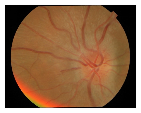
Swelling of the nerve, when the inflamed segment is anterior enough to be visible, produces the disc oedema seen on fundus examination in a minority of cases; more commonly the inflammation is retrobulbar, behind the visible portion of the nerve, and the disc appears entirely normal at presentation despite significant vision loss, the origin of the classic teaching that “the patient sees nothing and the doctor sees nothing.”
Fundus Explorer Pro
Photograph the retinal findings described here with the phone already in your pocket — 22 D optics and built-in illumination in one handheld unit.
From Choroida — the team behind this siteClinical Presentation
Subacute, progressive unilateral vision loss developing over hours to days is typical, often reaching its worst point within one to two weeks before beginning to improve.
Pain, particularly with eye movement, is present in the large majority of cases and is often the symptom that prompts the patient to seek attention before the vision loss becomes severe.
Colour desaturation, described by patients as colours looking washed out or less vivid in the affected eye, is a sensitive early sign, and a relative afferent pupillary defect is virtually always present in unilateral cases, making pupil testing one of the most useful bedside findings in this condition.
Examination Findings
A relative afferent pupillary defect is the single most reliable objective sign, present in essentially every case of unilateral optic neuritis regardless of how the disc itself looks.
The optic disc appears swollen in a minority of cases, described as papillitis, while the majority show a normal-appearing disc at presentation, reflecting retrobulbar inflammation. Visual field testing typically shows a central or cecocentral scotoma, though other patterns of field loss can occur.
Diagnostic Evaluation
MRI of the brain and orbits with contrast is the key investigation, both to confirm optic nerve enhancement supporting the diagnosis and, critically, to assess for white matter lesions elsewhere in the brain, since the number and pattern of these lesions is the strongest predictor of subsequent conversion to multiple sclerosis.
Aquaporin-4 antibody testing and MOG antibody testing should be considered, particularly in atypical presentations, since a positive result changes both the treatment approach and the long-term prognosis substantially compared with typical demyelinating optic neuritis.
Optical coherence tomography of the retinal nerve fibre layer, while not required for acute diagnosis, is useful for documenting baseline thickness and tracking the atrophy that often follows resolution of the acute episode.
Differential Diagnosis
- Non-arteritic anterior ischaemic optic neuropathy, typically painless, in an older patient with vascular risk factors, and with disc swelling that is usually segmental rather than diffuse
- Arteritic anterior ischaemic optic neuropathy from giant cell arteritis, an emergency requiring urgent recognition given the risk to the fellow eye without prompt corticosteroid treatment
- Compressive optic neuropathy from a tumour, which tends to be gradual and progressive rather than subacute, and typically painless
- Toxic or nutritional optic neuropathy, usually bilateral, gradual in onset, and associated with a relevant exposure or deficiency history
- Leber hereditary optic neuropathy, painless, often sequentially bilateral, and confirmed with mitochondrial genetic testing
Pain with eye movement and the subacute time course are what most reliably distinguish typical optic neuritis from ischaemic and compressive causes, which explains why these two features carry so much diagnostic weight at the initial visit.
Management
Intravenous corticosteroids, typically high-dose methylprednisolone, speed visual recovery but do not change the final visual outcome, which is why treatment is generally reserved for more severe cases or when faster recovery is clinically important, rather than given automatically to every patient.
Oral corticosteroids alone, without preceding intravenous treatment, are specifically avoided, since the Optic Neuritis Treatment Trial found an increased rate of recurrence with this approach compared with intravenous treatment or observation.
Disease-modifying therapy for multiple sclerosis is initiated based on MRI findings and overall risk of conversion, in coordination with neurology, and this decision is generally more consequential for long-term outcome than any treatment decision made for the optic neuritis episode itself.
Prognosis
Visual recovery in typical optic neuritis is generally good, with the large majority of patients regaining vision close to their baseline over weeks to months, even without treatment, though subtle deficits in colour vision or contrast sensitivity often persist even after acuity has returned to normal.
The risk of progression to multiple sclerosis after a first episode is directly related to the burden of white matter lesions seen on baseline MRI, ranging from a low risk with a normal scan to a substantially higher risk when multiple lesions are present, which is why MRI findings, more than any clinical feature, drive both prognosis counselling and the decision to start disease-modifying therapy.
Atypical optic neuritis, particularly when related to neuromyelitis optica spectrum disorder or MOG antibody disease, carries a distinctly different prognosis, with a greater risk of poor visual recovery and recurrence, reinforcing why identifying atypical features early changes both treatment and the conversation with the patient about what to expect.


Document what you see
Two smartphone imaging tools built for everyday clinic use — one for the slit lamp, one for the fundus.
From Choroida — the team behind this siteReferences
- Beck RW, Cleary PA, Anderson MM, et al. A randomized, controlled trial of corticosteroids in the treatment of acute optic neuritis (Optic Neuritis Treatment Trial). New England Journal of Medicine. 1992.
- Optic Neuritis Study Group. Multiple sclerosis risk after optic neuritis: final Optic Neuritis Treatment Trial follow-up. Archives of Neurology. 2008.
- Petzold A, Wattjes MP, Costello F, et al. The investigation of acute optic neuritis: a review and proposed protocol. Nature Reviews Neurology. 2014.
- Optic Neuritis. EyeWiki, American Academy of Ophthalmology.
- Optic Neuritis. StatPearls, NCBI Bookshelf.
Test yourself
A few questions straight from this article.
-
Which presentation characterises typical demyelinating optic neuritis?
The typical form studied in the Optic Neuritis Treatment Trial is unilateral, painful on eye movement, and generally recovers well regardless of whether it is treated. -
Why does the optic disc look normal in most patients presenting with optic neuritis?
Only an anteriorly placed inflamed segment produces visible disc oedema; more commonly the process is retrobulbar, so the patient sees nothing and the doctor sees nothing. -
Which objective sign is present in essentially every case of unilateral optic neuritis?
The relative afferent pupillary defect is the single most reliable objective sign, present whatever the disc itself looks like, which makes pupil testing the key bedside manoeuvre. -
What visual outcome is expected after a first episode of typical optic neuritis?
The large majority recover close to baseline acuity over weeks to months even without treatment, though colour vision and contrast sensitivity deficits often persist. -
Which visual field defect is typical in optic neuritis?
Central or cecocentral scotoma is the usual pattern on field testing, although other patterns of field loss can occur. -
Which baseline finding most strongly predicts conversion to multiple sclerosis after optic neuritis?
Risk rises directly with the number of white matter lesions on baseline MRI, from low with a normal scan to substantially higher with multiple lesions, driving both counselling and disease-modifying therapy. -
Which tests should be considered when optic neuritis presents with atypical features such as bilateral involvement?
A positive aquaporin-4 or MOG antibody result substantially changes both treatment and long-term prognosis compared with typical demyelinating optic neuritis. -
Why are oral corticosteroids alone avoided in treating optic neuritis?
The Optic Neuritis Treatment Trial found a higher recurrence rate with oral steroids alone compared with intravenous treatment or simple observation. -
What does intravenous methylprednisolone achieve in acute optic neuritis?
Because the final visual result is unchanged, intravenous steroids are generally reserved for more severe cases or when faster recovery matters clinically. -
Which feature best separates non-arteritic anterior ischaemic optic neuropathy from typical optic neuritis?
Pain with eye movement and the subacute time course are what most reliably distinguish optic neuritis from ischaemic causes, which are typically painless with segmental disc swelling.

Writes and reviews the clinical articles on Educate. Every post is checked against peer-reviewed sources before it goes live.


