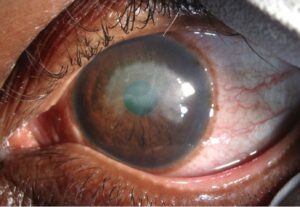
Ocular brucellosis is an uncommon but well-described complication of systemic infection with Brucella species, most often Brucella melitensis.
It causes uveitis in its many forms, and it can involve the retina, choroid, optic nerve, and cornea.
In regions where brucellosis is endemic, including the Middle East, the Mediterranean, and parts of Africa and Latin America, it belongs on the list of causes of unexplained uveitis.
The diagnosis rests on suspicion, serology, and a history of exposure to unpasteurized dairy products or infected livestock.
What Is Ocular Brucellosis?
Brucellosis is a zoonosis acquired by ingestion of unpasteurized milk or cheese, contact with infected animal tissue, or inhalation of aerosols.
It is one of the most common bacterial zoonoses worldwide.
Systemic disease produces undulating fever, night sweats, arthralgia, fatigue, and weight loss, and it may involve bone, joints, liver, spleen, and the nervous system.
Ocular involvement occurs in a small proportion of patients and may appear during the acute illness, during relapse, or as an isolated late manifestation.
Some patients present with uveitis without a clear history of febrile illness.
Ocular Manifestations
Brucella can affect almost every part of the eye.
Uveitis
Uveitis is the most frequent ocular manifestation.
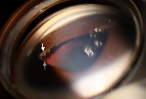
- Anterior uveitis, often granulomatous with mutton-fat keratic precipitates, or nongranulomatous
- Intermediate uveitis with vitritis and snowbanking
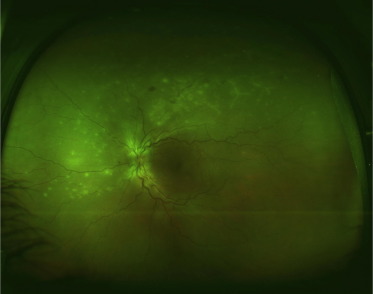
- Posterior uveitis with multifocal choroiditis
- Panuveitis
The course may be acute or chronic, and recurrences are common.
Retina and Choroid
- Multifocal choroiditis with round yellowish infiltrates
- Retinal vasculitis with sheathing and hemorrhage
- Macular edema
- Endogenous endophthalmitis in rare, severe cases
Optic Nerve
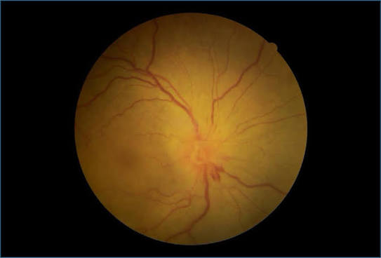
Optic neuritis, papillitis, and papilledema occur, particularly in neurobrucellosis (see papilledema).
Cornea and Sclera
Nummular keratitis and scleritis are reported.
Ocular brucellosis is therefore a possible cause of infectious uveitis in any of these patterns, and it should be considered next to tuberculosis and syphilis (see ocular tuberculosis and ocular syphilis).
Fundus Explorer Pro
Photograph the retinal findings described here with the phone already in your pocket — 22 D optics and built-in illumination in one handheld unit.
From Choroida — the team behind this siteDiagnosis
History and Systemic Assessment
Ask about consumption of raw milk or soft cheese, contact with sheep, goats, or cattle, occupational exposure such as farming or slaughterhouse work, travel to endemic regions, and previous fever or joint pain.
Systemic examination may reveal hepatosplenomegaly, lymphadenopathy, arthritis, or spinal tenderness.
Laboratory Tests
- Serology: the Rose Bengal test is a rapid screening test, and the standard tube agglutination test is commonly used, with a titer of 1:160 or higher considered significant in endemic regions
- Coombs test for brucella agglutinins, useful in chronic disease
- Blood culture, which is positive in a proportion of acute cases and requires prolonged incubation
- PCR on blood, aqueous, or vitreous when available
- ELISA for IgM and IgG
Serology may be negative in localized ocular disease, and a high index of suspicion is needed.
Other causes of uveitis should be excluded, including tuberculosis, syphilis, toxoplasmosis, and sarcoidosis, and testing should be chosen according to the clinical pattern.
Imaging
Fluorescein angiography identifies vasculitis and choroidal lesions.
OCT shows macular edema and subretinal fluid.
Systemic imaging is directed by symptoms, for example for spinal or hepatosplenic disease.
Differential Diagnosis
- Tuberculous uveitis
- Syphilitic uveitis
- Toxoplasma retinochoroiditis (see ocular toxoplasmosis)
- Sarcoid uveitis
- Behçet disease
- Bartonella neuroretinitis
- Lymphoma and other masquerade syndromes
Chronic granulomatous uveitis of unknown cause in an endemic region should lead to a specific search for Brucella.
What to Ask in a Patient With Uveitis in an Endemic Area
A short structured history often finds the diagnosis before any test is ordered.
- Any fever lasting weeks, especially with night sweats or a pattern of waxing and waning temperature
- Joint, back, or testicular pain
- Consumption of raw milk, fresh cheese, or undercooked liver
- Work with livestock, veterinary work, or laboratory exposure
- Family members with a similar illness, since exposure is often shared through food
Patients who have already received a partial course of antibiotics for another reason may have negative cultures, so a negative culture does not close the question.
Uveitis that relapses each time steroids are tapered, in a patient without a systemic disease label, is another cue to reconsider infection.
Treatment
Antibiotic Therapy
Treatment combines antibiotics that penetrate cells and can be given for prolonged periods.
- The World Health Organization regimen is doxycycline plus rifampin for six weeks
- Doxycycline for six weeks combined with streptomycin or gentamicin for the first two to three weeks is an alternative regimen with a lower relapse rate in many series
- Neurobrucellosis and severe ocular disease may need triple therapy and longer treatment, often with ceftriaxone in the initial phase
Ocular disease is often treated for a longer period than uncomplicated systemic infection, and duration is guided by clinical response and inflammatory markers.
Anti-Inflammatory Treatment
Topical corticosteroids and cycloplegics are used for anterior uveitis.
Systemic or periocular corticosteroids may be added once antibiotics are under way and infection is controlled, to reduce the damage caused by inflammation.
Steroids should not be given alone, because they can worsen infection.
Relapse and Follow-Up
Relapse occurs in a minority of patients, usually within months of stopping therapy, and it is more likely after short or single-drug courses.
Follow-up combines clinical examination, serology, and inflammatory markers, and involvement of infectious disease physicians is important.
Public health measures include pasteurization of dairy products and control of infection in livestock.
Prognosis
With timely antibiotics, most patients recover good vision.
Delayed diagnosis leads to chronic inflammation, macular edema, optic atrophy, cataract, and glaucoma.
Awareness in endemic areas reduces diagnostic delay.


Document what you see
Two smartphone imaging tools built for everyday clinic use — one for the slit lamp, one for the fundus.
From Choroida — the team behind this siteReferences
- Pappas G, Akritidis N, Bosilkovski M, Tsianos E. Brucellosis. N Engl J Med. 2005;352:2325-2336.
- Rolando I, Olarte L, Vilchez G, et al. Ocular manifestations associated with brucellosis: a 26-year experience in Peru. Clin Infect Dis. 2008;46:1338-1345.
- Ariza J, Bosilkovski M, Cascio A, et al. Perspectives for the treatment of brucellosis in the 21st century: the Ioannina recommendations. PLoS Med. 2007;4:e317.
- Corbel MJ. Brucellosis in Humans and Animals. Geneva: World Health Organization; 2006.
Test yourself
A few questions straight from this article.
-
Which Brucella species most often causes ocular brucellosis?
Ocular brucellosis is an uncommon but well-described complication of systemic infection with Brucella species, most often Brucella melitensis. -
Which history should raise suspicion of brucellosis in a patient with unexplained uveitis?
Brucellosis is a zoonosis acquired from unpasteurized milk or cheese, contact with infected animal tissue, or inhaled aerosols, so dietary and occupational exposure must be asked about directly. -
What is the most frequent ocular manifestation of brucellosis?
Uveitis is the most frequent ocular manifestation and may be anterior, intermediate, posterior or panuveitis, with an acute or chronic course and frequent recurrences. -
Which anterior segment picture is typical of brucellar anterior uveitis?
Brucellar anterior uveitis is often granulomatous with mutton-fat keratic precipitates, although a nongranulomatous pattern also occurs. -
Which corneal finding is reported in ocular brucellosis?
Nummular keratitis and scleritis are both reported in ocular brucellosis, alongside uveitis, chorioretinal and optic nerve involvement. -
Which serum tube agglutination titre is considered significant for brucellosis in endemic regions?
The standard tube agglutination test is commonly used, with a titre of 1:160 or higher considered significant in endemic regions, while the Rose Bengal test serves as a rapid screen. -
What does negative brucella serology mean in a patient with suspected ocular brucellosis?
Serology may be negative in localized ocular disease, and a negative culture does not close the question either, particularly after a partial course of antibiotics. -
What is the World Health Organization antibiotic regimen for brucellosis?
The World Health Organization regimen is doxycycline plus rifampin for six weeks; doxycycline with streptomycin or gentamicin for the first two to three weeks is an alternative with a lower relapse rate in many series. -
Why must corticosteroids never be used alone in ocular brucellosis?
Steroids should not be given alone because they can worsen the infection; systemic or periocular steroid is added only once antibiotics are under way and infection is controlled. -
What is the visual outcome of ocular brucellosis?
With timely antibiotics most patients recover good vision, whereas delayed diagnosis leads to chronic inflammation, macular edema, optic atrophy, cataract and glaucoma.

Writes and reviews the clinical articles on Educate. Every post is checked against peer-reviewed sources before it goes live.