
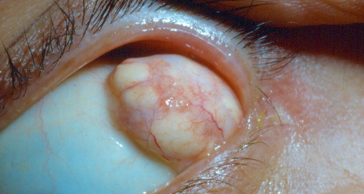
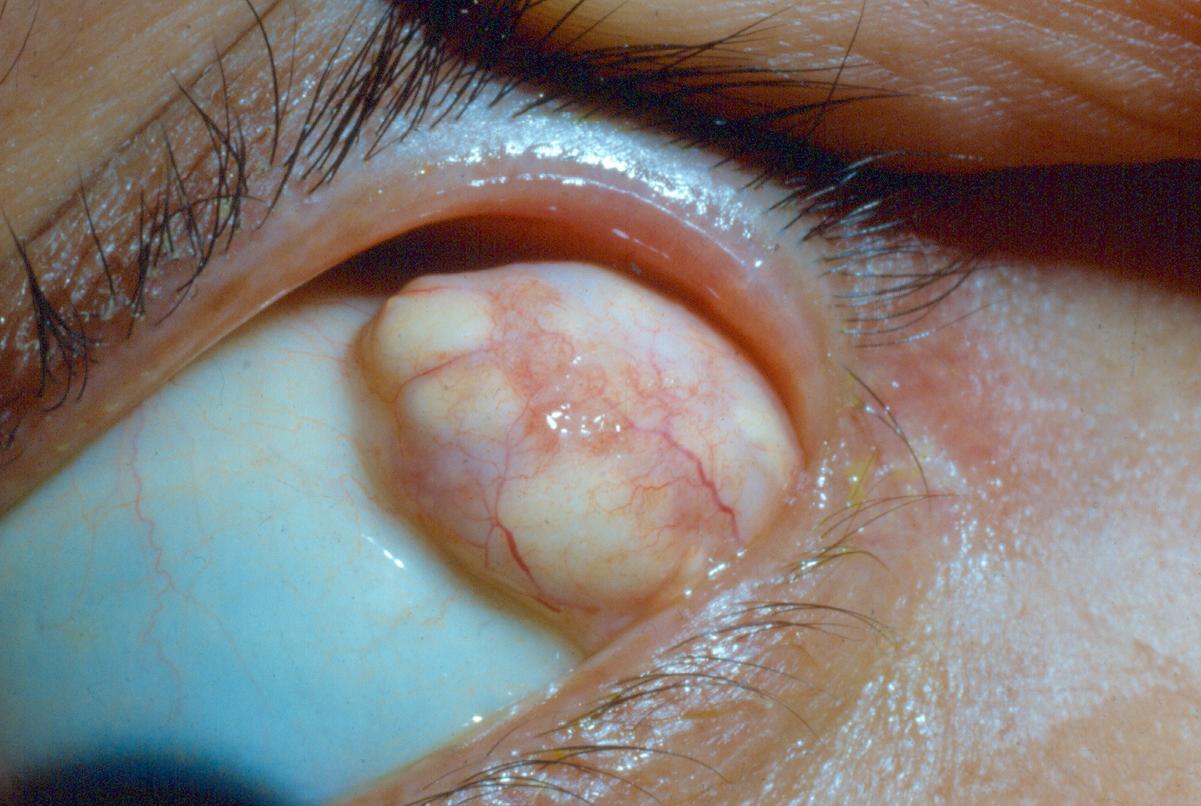
Orbital dermoid cysts are benign congenital choristomas — masses composed of tissue that is normal in itself but abnormal in location — arising from ectodermal tissue trapped along embryonic bony suture lines during development, and they represent one of the most common orbital masses encountered in children.
Their typically slow, indolent growth and highly characteristic imaging appearance mean that in the great majority of cases, the diagnosis can be made with real confidence well before any tissue is obtained, which directly shapes how conservatively or aggressively they are managed.
Pathogenesis
During facial and orbital development, ectodermal (and sometimes mesodermal) tissue can become trapped along the lines where adjacent bones fuse, most commonly at the frontozygomatic suture in the superotemporal orbit or, less commonly, along the superonasal orbital rim.
This trapped tissue continues to differentiate normally into skin and skin appendage elements — hair follicles, sebaceous glands, sweat glands — but does so in an abnormal, deep location, producing a cyst that gradually fills with the desquamated keratin and sebaceous material these normally functioning skin elements continuously produce.
Clinical Presentation
Most orbital dermoid cysts present in infancy or early childhood as a firm, painless, slowly enlarging mass at the superotemporal or, less often, superonasal orbital rim, often noted incidentally by a parent rather than causing any functional visual symptoms, because the great majority remain small and superficial without significantly affecting the globe or optic nerve.
A subset, however, extend more deeply into the orbit along the suture line, and these deeper dermoids can present later, sometimes not until adolescence or adulthood, with proptosis or displacement of the globe as they slowly enlarge over years.
All-fit Slit-Lamp Adapter
Record and share exactly what you see at the slit lamp. One adapter fits any slit lamp or surgical microscope — and any smartphone.
From Choroida — the team behind this siteExam Findings
- A firm, smooth, mobile or minimally mobile subcutaneous mass, most commonly at the superotemporal orbital rim, generally non-tender unless the cyst has ruptured (see below)
- Normal overlying skin, without inflammation, in an uncomplicated, unruptured cyst
- Deeper orbital dermoids may produce proptosis, globe displacement, or, rarely, optic nerve compression if large enough and positioned to affect these structures
- Some dermoids have a dumbbell configuration, with a superficial component and a separate, connected deep intraorbital or even intracranial component communicating through a bony defect along the suture line — a configuration with real implications for surgical planning, since incomplete recognition of a deep component can lead to incomplete excision and recurrence
Complication: Rupture
A dermoid cyst that ruptures, whether spontaneously or from trauma, releases its keratin and sebaceous contents into surrounding orbital tissue, provoking an intense granulomatous inflammatory response that produces acute pain, redness, and swelling — a presentation that can closely mimic orbital cellulitis or an acutely inflamed process.
Recognizing a ruptured dermoid as the actual cause (rather than an infectious process) matters directly for appropriate management, which centers on complete surgical removal of the cyst and its released contents rather than antibiotics alone.
Differential Diagnosis
- Epidermoid cyst — a related choristoma lacking the skin appendage (hair follicle, sebaceous gland) elements of a true dermoid, generally indistinguishable clinically and managed similarly
- Capillary hemangioma — typically presents somewhat differently, with a bluish or reddish color if superficial and a history of growth in early infancy followed by later involution, discussed in its own dedicated article on this site
- Lymphangioma — generally less well-circumscribed, can fluctuate in size (sometimes acutely, with intralesional hemorrhage), and has its own characteristic imaging appearance, discussed in its own dedicated article on this site
- Encephalocele — a herniation of intracranial contents through a bony defect, an important and more serious consideration for masses in the superonasal location specifically, given the proximity to the anterior cranial fossa; imaging is essential to exclude this before any surgical intervention is attempted, since inadvertently entering an encephalocele during what was presumed to be simple dermoid excision would be a serious complication
Diagnostic Evaluation
CT or MRI is obtained for any dermoid with imaging features suggesting deep extension, a dumbbell configuration, or proximity to the orbital rim bone in a location (superonasal) where an encephalocele needs to be excluded.
Imaging characteristically shows a well-circumscribed cystic lesion, often with a smooth, scalloped remodeling of the adjacent bone reflecting its slow, longstanding growth pattern.
A small, superficial dermoid with classic clinical features and no concerning imaging red flags may reasonably proceed to excision without extensive preoperative imaging, though many surgeons favor at least baseline imaging to confirm the extent of the lesion before planning surgery.
Management
Complete surgical excision is the definitive treatment and is generally recommended even for small, asymptomatic dermoids, both to prevent the complication of rupture (with its associated acute inflammatory presentation) and to address the cosmetic concern most families have about a visible mass, and careful, complete removal of the entire cyst wall is important to prevent recurrence.
For dermoids with a deep or dumbbell configuration, more extensive surgical planning — sometimes involving neurosurgical co-management when there is any suspicion of intracranial extension or communication — is warranted rather than proceeding with a straightforward, limited excision as would be appropriate for a purely superficial lesion, since attempting to remove a deep component without adequate exposure risks incomplete excision and eventual recurrence.


Document what you see
Two smartphone imaging tools built for everyday clinic use — one for the slit lamp, one for the fundus.
From Choroida — the team behind this siteReferences
- Bonavolontà P, Dell’Aversana Orabona G, Abbate V, et al. Orbital dermoid cyst: case series and review of the literature. Journal of Craniofacial Surgery.
- Shields JA, Kaden IH, Eagle RC Jr, Shields CL. Orbital dermoid cysts: clinicopathologic correlations, classification, and management. Ophthalmic Plastic and Reconstructive Surgery.
- American Academy of Ophthalmology. Basic and Clinical Science Course, Section 6: Pediatric Ophthalmology and Strabismus.
