Eyelid lumps are common and usually harmless.
A small number are skin cancers that threaten the eye.
Knowing the warning signs avoids missed malignancy.
Common Benign Eyelid Lumps
Chalazia, styes, cysts and papillomas make up most lesions.
They are smooth, mobile and slow growing.
Pain suggests infection.
Therefore, history and shape guide the diagnosis (see chalazion).
Stye and Chalazion
A stye is a tender infected gland.
A chalazion is a painless granuloma from a blocked gland (see hordeolum and stye).
Both respond to warm compresses.
So treat first and watch the course.
All-fit Slit-Lamp Adapter
Record and share exactly what you see at the slit lamp. One adapter fits any slit lamp or surgical microscope — and any smartphone.
From Choroida — the team behind this siteCysts Causing Eyelid Lumps
Moll gland cysts are clear and translucent.
Zeis gland cysts are small and yellowish.
Epidermoid cysts are firm and white.
Also these lesions can be excised if bothersome.
Xanthelasma
Yellowish plaques appear on the medial lids.
They relate to lipid disorders in some patients (see xanthelasma).
Check lipid levels.
Therefore, refer for systemic evaluation.
Papilloma and Skin Tag Eyelid Lumps
Papillomas appear as fleshy stalks.
Skin tags are common in older adults.
Removal is simple.
So reassure patients about their benign nature.
Molluscum Eyelid Lumps
Molluscum contagiosum causes dome-shaped umbilicated papules.
It may cause conjunctivitis from shedding virus.
Treatment includes curettage.
Also look at the conjunctiva.
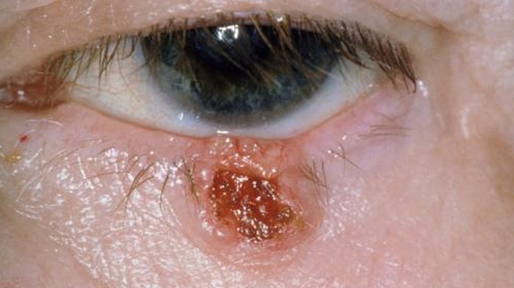
Basal Cell Carcinoma
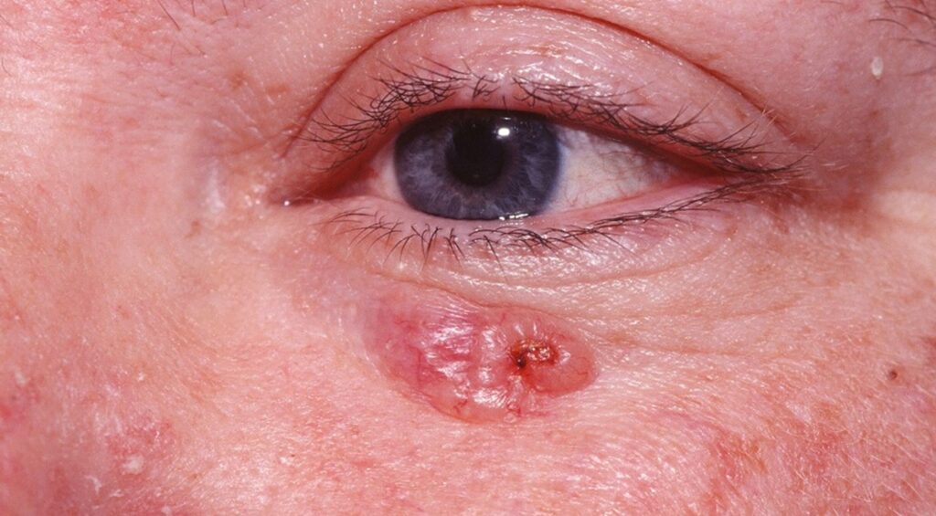
This is the commonest malignant eyelid tumor.
It often appears as a pearly nodule with telangiectasia (see basal cell carcinoma of the eyelid).
The lower lid and medial canthus are common sites.
So biopsy suspicious lesions.
Squamous Carcinoma Eyelid Lumps
Squamous carcinoma presents as a scaly, ulcerated nodule.
It can spread along nerves.
Sun exposure is a risk factor.
Therefore, refer early.
Sebaceous Carcinoma
This tumor can mimic chronic chalazion or blepharitis.
Loss of lashes and persistent thickening are clues.
Biopsy is essential.
Also look for pagetoid spread.
Melanoma
Pigmented lesions that change need review.
Irregular borders and growth are worrying.
Biopsy confirms the diagnosis.
So photograph and monitor naevi.
Red Flags
Loss of lashes, ulceration, bleeding and rapid growth suggest cancer.
A lesion that recurs after treatment also needs biopsy.
Asymmetry and distortion of the lid margin raise concern.
Therefore, send tissue for pathology.
Management
Benign lesions can be observed or removed for comfort.
Malignant ones need excision with margins and reconstruction (see periocular basal cell carcinoma reconstruction).
Mohs surgery is used in some cases.
So refer to an oculoplastic surgeon.
Eyelid Lumps in Children
Chalazia and molluscum are common.
Rare tumors occur.
Persistent lesions need review.
Also assess for amblyopia if the lid droops.
Eyelid Lumps: Practical Summary
Look at shape, surface, lashes and course.
If features worry you, biopsy.
Because early removal is easier, do not wait.
Finally, advise sun protection.
Pitfalls to Avoid
- Do not assume every lump is a chalazion
- Never send a recurrent lesion away without pathology
- Avoid ignoring loss of lashes
- Do not forget to examine the conjunctiva
Documentation of Eyelid Lumps
Record size, color, surface and margin involvement.
Take a photograph with a ruler.
Because change over time matters, compare images.
Therefore, review lesions at set intervals.
Biopsy Technique
Incisional or excisional biopsy depends on size and site.
Take tissue from the edge and the center of a large lesion.
Because margins matter for cancer, label the specimen.
So send the sample for pathology.
Frozen Section and Mohs Surgery
Frozen sections check margins during surgery.
Mohs surgery spares tissue and gives high cure rates.
Because the eyelid is delicate, this saves function.
Therefore, refer to a team with experience.
Reconstruction
After removal of a tumor, the lid needs reconstruction.
Grafts and flaps restore the margin.
Because function matters, protect the eye.
So plan reconstruction before surgery.
Follow-Up After Treatment
Review the site for recurrence.
Check regional lymph nodes in aggressive tumors.
Because new tumors can arise, examine the skin.
Finally, advise sun protection.
Patient Questions
Patients ask whether a lump will become cancer.
Most do not, but monitoring helps.
They also ask whether removal leaves a scar.
Careful technique keeps scars small.
Eyelid Lumps: Practical Summary
When a lump has been present for weeks, examine the lash line and the margin.
If you suspect cancer, biopsy without delay.
Because most lumps are benign, reassure patients when the features are typical.
Finally, refer all lesions that change.
Eyelid Lumps: Sun Protection
Wear a hat and sunglasses outdoors.
Apply sunscreen to the skin around the eye with care.
Because ultraviolet light drives skin cancer, protection works.
Therefore, make it part of routine advice.
For the latest studies, see the PubMed results for eyelid lumps.


Document what you see
Two smartphone imaging tools built for everyday clinic use — one for the slit lamp, one for the fundus.
From Choroida — the team behind this siteReferences
- Cook BE Jr, Bartley GB. Epidemiologic characteristics and clinical course of patients with malignant eyelid tumors in an incidence cohort in Olmsted County, Minnesota. Ophthalmology. 1999;106:746-750.
- Slutsky JB, Jones EC. Periocular cutaneous malignancies: a review of the literature. Dermatol Surg. 2012;38:552-569.
- Yin VT, Merritt HA, Sniegowski M, Esmaeli B. Eyelid and ocular surface carcinoma: diagnosis and management. Clin Dermatol. 2015;33:159-169.
- Shields JA, Shields CL. Eyelid, Conjunctival, and Orbital Tumors: An Atlas and Textbook. 3rd ed. Wolters Kluwer; 2016.

Writes and reviews the clinical articles on Educate. Every post is checked against peer-reviewed sources before it goes live.