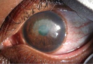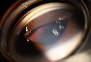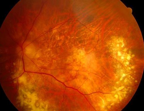
Candle wax drippings, or taches de bougie, are one of the more recognizable retinal findings in ocular sarcoidosis.
Along the retinal veins, usually in the periphery, you see yellow-white, globular, perivenous infiltrates that look like wax that has run down the side of a candle and hardened.
What causes the appearance
The material is a perivascular cuff of inflammatory cells and exudate that accumulates around inflamed veins.
In sarcoid it is granulomatous, and the cuffing is patchy, not continuous, which gives the knobby, irregular outline.
Small hemorrhages, vascular leakage, and occasionally neovascularization can follow.
When you will see it
- Sarcoidosis is the most typical setting (see ocular sarcoidosis and sarcoid uveitis)
- Tuberculosis and other granulomatous infections
- Multiple sclerosis-associated intermediate uveitis, where periphlebitis is a feature
- Pars planitis, often with snowbanking and snowballs in the vitreous (see pars planitis)
- Behçet disease, in which periphlebitis tends to be occlusive and more severe
- Eales disease and other peripheral retinal vasculopathies
The periphlebitis of sarcoid can occur with little or no anterior chamber inflammation, so a quiet front of the eye does not exclude it.
Related findings that support sarcoid
- Mutton-fat keratic precipitates and iris nodules (see keratic precipitates)
- “String-of-pearls” vitreous opacities
- Optic disc granuloma or swelling
- Choroidal granulomas and, in some cases, macular edema
- Bilateral hilar lymphadenopathy on chest imaging
Investigation
- Chest imaging, ideally a high-resolution CT, since chest radiographs miss many cases
- Serum angiotensin-converting enzyme and lysozyme, which are supportive but neither sensitive nor specific
- Tuberculosis testing before steroid or immunosuppressive treatment
- Fluorescein angiography to show leakage along the veins, capillary non-perfusion, and macular edema
- Biopsy of accessible lesions, when the diagnosis is uncertain
Treatment
Periphlebitis without macular edema or vitritis may need no treatment beyond observation.
When there is significant inflammation or macular edema, treatment typically follows a stepwise path.
- Periocular or intravitreal corticosteroid injection for unilateral disease
- Oral corticosteroids for bilateral or severe disease, with a taper plan
- Steroid-sparing agents such as methotrexate or mycophenolate when long-term control is needed
- Adalimumab in refractory cases
- Laser or anti-VEGF for retinal neovascularization after ischemia is documented
Looking for it in practice
The sheathing tends to start in the peripheral retina, so the finding is easily missed without indirect ophthalmoscopy and scleral depression.
Many patients are seen with vague floaters and blurred vision, and a dilated exam with a wide field of view is the only way to find it.
Wide-field fluorescein angiography is helpful because it shows vascular leakage and capillary dropout that are invisible on clinical examination, and it can show how much of the disease is active when the clinical picture is mixed.
A practical point is that sheathing should be differentiated from the inactive, white, narrowed vessel sleeves of old inflammation.
Active periphlebitis is fluffy and yellow, and it is associated with leakage on angiography.
Inactive sheathing is thin, white, and without leakage.
That distinction decides whether treatment needs to be escalated, since inactive sheathing is a scar and does not need treatment (see retinal vasculitis sheathing patterns).
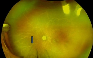
Distinguishing active periphlebitis from old sheathing
Active periphlebitis is fluffy, yellow-white, and associated with leakage on fluorescein angiography and with cells in the vitreous. Old sheathing is thin, white, and sharply demarcated, without leakage or vitritis. The distinction matters because inactive sheathing is a scar and needs no treatment, while active periphlebitis can lead to occlusion, ischemia, and neovascularization.
Following the patient
Wide-field fluorescein angiography helps to document leakage and nonperfusion in the periphery, and it can guide laser to ischemic retina if neovascularization develops. Follow the patient with serial examinations and imaging while treatment is tapered. Because sarcoidosis is a systemic disease, coordinate with a physician for chest imaging, pulmonary function, and monitoring of the effects of long-term immunosuppression.
Pitfalls
- Do not exclude sarcoid because the front of the eye is quiet
- Do not rely on a normal chest radiograph, since high-resolution CT is more sensitive
- Do not start steroids before tuberculosis and syphilis have been considered
- Do not forget that periphlebitis may be the first sign of multiple sclerosis in a young adult
What to tell the patient
Explain that the retinal changes reflect inflammation of the vessel walls, that treatment is aimed at calming it, and that regular examinations are needed because the disease can come back. Encourage them to report new floaters, blurred vision, or pain.
Prognosis
Many patients do well, but sarcoid can relapse years later.
Long-term follow-up with attention to intraocular pressure, cataract, and recurrent macular edema is important.
Patients should also know that sarcoidosis is a systemic disease, and that a pulmonologist or rheumatologist should be involved in the care, because the eye may be the only organ showing symptoms at the time of diagnosis.


Document what you see
Two smartphone imaging tools built for everyday clinic use — one for the slit lamp, one for the fundus.
From Choroida — the team behind this siteReferences
- Herbort CP, Rao NA, Mochizuki M. International criteria for the diagnosis of ocular sarcoidosis: results of the first International Workshop on Ocular Sarcoidosis. Ocul Immunol Inflamm. 2009;17:160-169.
- Jabs DA, Johns CJ. Ocular involvement in chronic sarcoidosis. Am J Ophthalmol. 1986;102:297-301.
- Pasadhika S, Rosenbaum JT. Ocular sarcoidosis. Clin Chest Med. 2015;36:669-683.
- Dana MR, Merayo-Lloves J, Schaumberg DA, Foster CS. Prognosticators for visual outcome in sarcoid uveitis. Ophthalmology. 1996;103:1846-1853.
Test yourself
A few questions straight from this article.
-
What do candle wax drippings look like on fundus examination?
Also called taches de bougie, they are yellow-white globular perivenous infiltrates, usually peripheral, resembling wax that has run down a candle and hardened. -
What is the material that forms candle wax drippings?
The cuff of inflammatory cells and exudate around inflamed veins is granulomatous in sarcoid, and because the cuffing is patchy rather than continuous it looks knobby and irregular. -
In which condition are candle wax drippings most typically seen?
Sarcoidosis is the most typical setting, although tuberculosis and other granulomatous infections, multiple sclerosis-associated intermediate uveitis, pars planitis, Behcet disease and Eales disease can also produce periphlebitis. -
How does the periphlebitis of Behcet disease tend to differ from that of sarcoidosis?
In Behcet disease the periphlebitis tends to be occlusive and more severe than the patchy granulomatous cuffing seen in sarcoid. -
What does a quiet anterior chamber mean in a patient with suspected sarcoid periphlebitis?
Sarcoid periphlebitis can occur with little or no anterior chamber inflammation, so a quiet front of the eye does not exclude it. -
Which features indicate that perivenous sheathing is active rather than an old scar?
Active periphlebitis is fluffy and yellow-white with angiographic leakage and vitreous cells, whereas inactive sheathing is thin, white and non-leaking, and being a scar needs no treatment. -
Which chest investigation is preferred when sarcoid periphlebitis is suspected?
High-resolution CT is preferred because chest radiographs miss many cases; serum angiotensin-converting enzyme and lysozyme are supportive but neither sensitive nor specific. -
Which test should be done before starting steroid or immunosuppressive treatment for periphlebitis?
Tuberculosis testing is required before steroid or immunosuppressive treatment, since granulomatous infection can produce the same perivenous appearance. -
How is periphlebitis without macular oedema or vitritis managed?
Periphlebitis without macular oedema or vitritis may need nothing beyond observation; stepwise treatment is reserved for significant inflammation or macular oedema. -
What should long-term follow-up of sarcoid periphlebitis watch for?
Sarcoid can relapse years later, so long-term follow-up attends to intraocular pressure, cataract and recurrent macular oedema, with a physician involved for the systemic disease.

Writes and reviews the clinical articles on Educate. Every post is checked against peer-reviewed sources before it goes live.