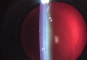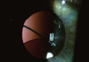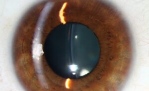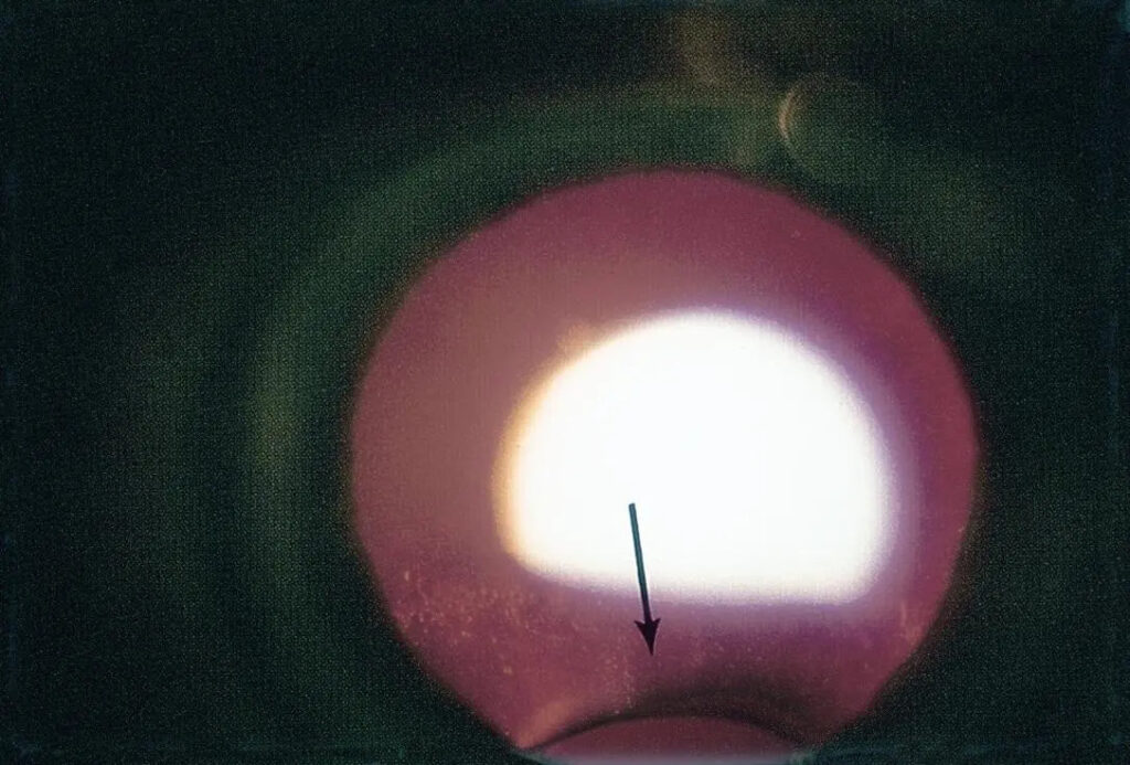
Lens coloboma is a congenital notch or flattening of the lens edge, usually inferior, where the zonular fibers are absent or deficient. It often sits beside an iris coloboma, yet the lens defect comes from missing zonular support rather than missing lens tissue. Vision is often good when the defect is small and isolated.
What Is a Lens Coloboma?
The word coloboma comes from the Greek for curtailed, and it describes a defect where tissue is missing or incomplete. In the iris, choroid, and optic disc, the cause is incomplete closure of the embryonic fissure. A lens coloboma differs: the lens equator looks notched because zonular fibers in that sector failed to develop.
The lens is not truly missing in that sector. The capsule and cortex are present, but the equator flattens where zonular tension is absent. Because the lens and iris share the same developmental field, the notch often lies in the same sector as an iris or choroidal coloboma, usually inferior or inferonasal.
How Coloboma of the Lens Looks on Examination
Dilate the pupil first. The notch appears as a flattened or scalloped segment of the equator, and the zonular fibers in that sector may be sparse or invisible at the slit lamp. Small defects are easy to miss, so scan the entire lens periphery deliberately.
Slit-lamp photographs typically show a dilated pupil with a straight or indented lens edge, often with an iris defect in the same quadrant. Retroillumination helps outline the contour. The defect can be unilateral or bilateral, and severity ranges from a subtle notch to a large indentation.
All-fit Slit-Lamp Adapter
Record and share exactly what you see at the slit lamp. One adapter fits any slit lamp or surgical microscope — and any smartphone.
From Choroida — the team behind this siteLens Coloboma Versus Iris Coloboma
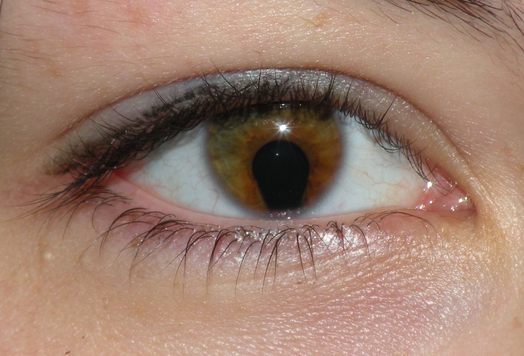
Iris coloboma is the more common finding, and it is what most patients picture when they hear coloboma of the eye. It results from incomplete closure of the embryonic fissure and leaves a gap in the iris, most often inferior. The pupil then takes a keyhole or teardrop shape, with the narrow end pointing down.
A star-shaped pupil is not typical of coloboma. Angular or irregular pupils more often come from trauma, sphincter tears, posterior synechiae, or prior surgery. A true iris coloboma has a sharply defined gap with no iris tissue in it, and it has been present since birth. Ask about injury or surgery before attributing a new shape to coloboma.
A Line or Notch in the Iris: What Else Could It Be?
A dark line or slit in the iris is not always coloboma. A surgical or laser iridotomy leaves a small, round or oval hole, usually superior. Iridodialysis, a traumatic tear at the iris root, creates a D-shaped pupil margin. A congenital coloboma usually sits inferiorly, is present from birth, and often accompanies other ocular anomalies. Slit-lamp examination separates these quickly.
Associated Findings
Examine the rest of the eye whenever you see a lens notch. Lens coloboma frequently travels with other structural defects, and the pattern of involvement guides both the systemic workup and the visual prognosis. Look specifically for the following:
- Iris coloboma, often in the matching sector
- Choroidal, retinal, and optic disc coloboma
- Cataract, sometimes related to the abnormal capsule and zonules in the affected area
- Microphthalmia, discussed in our workup of microphthalmia and coloboma syndromes
- Systemic syndromes such as CHARGE syndrome, especially when several ocular structures are involved
Genetic and Developmental Background
Lens coloboma is rare, and it is often found during the evaluation of a child with another coloboma. Many colobomatous eyes arise sporadically, though autosomal dominant, recessive, and X-linked patterns exist. Mutations in CHD7 cause CHARGE syndrome, so a child with multiple ocular colobomas warrants a systemic and genetic review.
Does a Lens Coloboma Affect Vision?
A small lens coloboma often causes little visual disturbance. Larger defects change lens curvature and can produce lenticular astigmatism or other irregular refractive error. Visual acuity also depends on associated findings, so a coexisting coloboma involving the macula or optic nerve, amblyopia, or cataract may limit vision far more than the lens notch.
The same zonular deficiency matters if the lens ever needs removal. A sector without zonules is a zone of reduced support during phacoemulsification, much like other causes of focal weakness described in lens subluxation. Plan ahead with capsular hooks or a capsular tension ring, and be prepared for a lens that shifts during capsulorhexis.
What Else Causes a Notched Lens?
A notched or displaced lens is not always a lens coloboma. Traumatic zonular dialysis, Marfan syndrome, homocystinuria, and microspherophakia all produce zonular weakness or abnormal lens shape. Unlike coloboma, these conditions often show lens shift, phacodonesis, or a small spherical lens, and they usually lack the matching iris or choroidal defect.
Diagnostic Evaluation
Diagnosis rests on a careful dilated examination, since the notch is easy to miss. Complete the workup in every affected child, because the lens finding rarely occurs alone and the rest of the eye often determines vision.
- Dilated slit-lamp examination of the full lens equator, with retroillumination
- Dilated fundus examination for choroidal, retinal, and optic disc coloboma
- Cycloplegic refraction to measure astigmatism and assess amblyopia risk in children
- Corneal diameter and axial length to look for microphthalmia
- Ultrasound biomicroscopy when the zonules and lens equator cannot be seen directly
- Referral for systemic and genetic evaluation when more than one ocular structure is involved, following the same principles used for any coloboma
Management of Lens Coloboma
Observation
Mild lens coloboma without visual impact or cataract needs observation only. The notch itself does not require treatment, and regular follow-up lets you track refraction, lens clarity, and any change in the notch over time. Children need particular attention so that amblyopia is caught early.
Correcting Refractive Error
Spectacles or contact lenses correct lenticular astigmatism and other refractive irregularity, as they do for irregular astigmatism from other causes. In children, correct significant refractive error early and treat amblyopia with patching or other standard therapy if it develops. Vision may be limited by posterior segment coloboma, so set expectations accordingly.
Cataract Surgery Considerations
Recognize the focal zonular weakness before surgery. Preoperative ultrasound biomicroscopy can help map the extent of the defect. Intraoperative tools include capsular hooks and a capsular tension ring or segment, and the surgeon should be ready for a scleral-fixated lens if capsular support proves inadequate. Warn the patient that outcomes depend on the extent of the zonular defect.
Prognosis
Isolated, mild lens coloboma generally carries a good visual prognosis. Management focuses on correcting refractive irregularity and watching for cataract over time. Visual outcome in the wider colobomatous eye depends more on whether the macula, optic nerve, or retina is involved than on the lens notch itself.
When the lens finding belongs to a broader pattern of colobomatous involvement, the care plan extends beyond the eye. Retinal detachment is a known risk in eyes with choroidal coloboma, and systemic associations need attention. A complete ocular and, when appropriate, systemic evaluation is warranted whenever a lens coloboma is identified.


Document what you see
Two smartphone imaging tools built for everyday clinic use — one for the slit lamp, one for the fundus.
From Choroida — the team behind this siteReferences
- Onwochei BC, Simon JW, Bateman JB, Couture KC, Mir E. Ocular colobomata. Surv Ophthalmol. 2000;45:175-194.
- Nelson LB, Maumenee IH. Ectopia lentis. Surv Ophthalmol. 1982;27:143-160.
- Pagon RA. Ocular coloboma. Surv Ophthalmol. 1981;25:223-236.
- Jesberg DO, Schepens CL. Retinal detachment associated with coloboma of the choroid. Arch Ophthalmol. 1961;65:163-173.
Test yourself
A few questions straight from this article.
-
What developmental abnormality produces a lens coloboma?
A lens coloboma arises from absent or deficient zonular fibres in a specific sector, which fail to give the lens equator the complete circumferential support it needs for a round contour. -
Incomplete closure of the fetal fissure accounts for coloboma of which structures?
Iris, choroidal and optic disc coloboma all follow incomplete fetal fissure closure, a mechanism distinct from the localized zonular deficiency behind a lens coloboma. -
What is the characteristic slit-lamp appearance of a lens coloboma?
The finding is a notch or flattened segment of the lens equator, best appreciated after pupillary dilation and easily overlooked unless specifically sought. -
Where is a lens coloboma most often located?
Lens colobomas are often sited inferiorly or inferonasally, although the exact position can vary between eyes. -
Which ocular coloboma most frequently coexists with a lens coloboma, often in a corresponding location?
A coexisting iris coloboma in the same quadrant is frequent, reflecting the shared developmental field from which these structures arise. -
Which systemic syndrome should be considered when lens coloboma coexists with iris or choroidal coloboma?
As with other ocular colobomas, systemic associations including CHARGE syndrome should be considered, particularly when several ocular structures are colobomatous. -
What refractive consequence can a larger lens coloboma produce?
Mild defects may disturb vision minimally, but larger ones can cause lenticular astigmatism or other refractive irregularity because the lens shape is altered. -
Why does a lens coloboma matter if cataract surgery is later required in that eye?
The localized absence of zonular support that created the coloboma is also a zone of reduced structural support at surgery, so planning may include capsular support devices. -
How is a mild lens coloboma without visual impact or cataract managed?
Without a visually significant complication the finding requires no direct treatment, so mild lens coloboma is simply observed. -
Which lens complication should be monitored for over time in an eye with lens coloboma?
Cataract can develop in association with a lens coloboma, sometimes related to the abnormal capsule and zonular support in the affected sector, so follow-up includes watching for it.

Writes and reviews the clinical articles on Educate. Every post is checked against peer-reviewed sources before it goes live.