
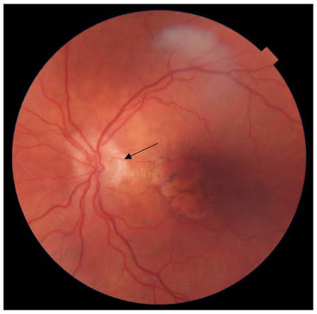
A cilioretinal artery is, for most of a person’s life, an entirely silent anatomic variant, noticed only incidentally on fundus examination or angiography, but its presence can become genuinely consequential in a single dramatic scenario: a central retinal artery occlusion, where this one additional vessel can be the difference between devastating, near-total vision loss and a meaningfully preserved island of central vision.
An Unusual Source of Blood Supply
Most of the retina’s blood supply comes from the central retinal artery, a branch of the ophthalmic artery, but in a meaningful proportion of the population, an additional vessel, the cilioretinal artery, supplies part of the retina, most often a portion of the macula or the area between the optic disc and macula, from an entirely different source: the posterior ciliary (choroidal) circulation rather than the central retinal artery system.
This distinct origin is the entire reason a cilioretinal artery matters clinically, since it means the territory it supplies has an independent blood source that is not affected when the central retinal artery itself becomes occluded.
Prevalence and Normal Appearance
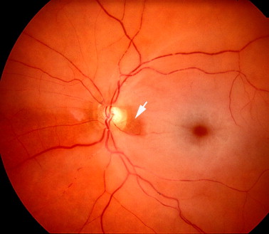
Cilioretinal arteries are present in a substantial minority of eyes, and when present, they typically emerge from the temporal border of the optic disc, coursing toward the macula, and are generally an incidental finding on routine fundus examination or fluorescein angiography, with no functional significance under normal circumstances.
Fundus Explorer Pro
Photograph the retinal findings described here with the phone already in your pocket — 22 D optics and built-in illumination in one handheld unit.
From Choroida — the team behind this siteClinical Significance in Central Retinal Artery Occlusion
When a central retinal artery occlusion occurs, the retinal territory normally supplied by the central retinal artery becomes acutely ischemic, typically producing severe, often near-total, vision loss.
- In an eye with a cilioretinal artery supplying part of the macula, that specific area continues to receive blood supply from the independent ciliary circulation even as the rest of the retina becomes ischemic from the central retinal artery occlusion, a phenomenon termed macular sparing
- This spared area can preserve a meaningful island of central visual function, sometimes allowing retention of good visual acuity despite severe, near-total peripheral field loss from the occlusion affecting the rest of the retina
- The specific location and extent of the cilioretinal artery’s territory determines how much, and which part, of central vision is preserved in a given affected patient
Clinical Recognition
- On fundus examination during an acute central retinal artery occlusion, the presence of a cilioretinal artery can sometimes be inferred from a localized area of relatively preserved, normal-appearing retina amid the surrounding ischemic, pale, edematous retina characteristic of the occlusion
- Fluorescein angiography can directly demonstrate the cilioretinal artery filling from the choroidal circulation, often notably earlier than the delayed or absent filling of the occluded central retinal artery territory, providing clear diagnostic confirmation of its presence and function
Other Clinical Contexts
A cilioretinal artery can occasionally itself become occluded, a cilioretinal artery occlusion, which produces a more limited, localized area of retinal ischemia corresponding specifically to its supply territory, generally with a correspondingly more limited visual field defect than a full central retinal artery occlusion, though the visual impact depends on whether the affected territory includes central, foveal retina.
Clinical and Prognostic Value
Recognizing the presence and territory of a cilioretinal artery in a patient who has experienced a central retinal artery occlusion provides genuinely useful prognostic information, helping explain, and anticipate, a better-than-expected degree of preserved central vision in an eye that would otherwise be expected to have very poor visual outcome following this typically devastating vascular event.
This anatomic variant is a clear illustration of how a normal, usually silent, developmental variation in blood supply can become the single most important factor determining visual outcome in an acute, sight-threatening vascular emergency.
Management
The cilioretinal artery itself requires no treatment, being a normal anatomic variant.
Management of central retinal artery occlusion follows standard urgent management principles for this ophthalmic emergency, and the presence of a cilioretinal artery, while influencing visual prognosis, does not change the urgency or overall approach to managing the underlying occlusive event and its associated systemic vascular risk factor workup.
A practical point in emergencies
A cilioretinal artery supplies part of the macula in a proportion of eyes, and when the central retinal artery is blocked, it may preserve central vision. Ask patients with sudden vision loss whether the center is spared. It should not delay urgent treatment, because the rest of the retina remains ischemic.
Prognosis
The presence of a functioning cilioretinal artery supplying macular territory is one of the most favorable prognostic factors in central retinal artery occlusion, often associated with meaningfully better preserved central visual acuity than would otherwise be expected from this typically severe vascular event.


Document what you see
Two smartphone imaging tools built for everyday clinic use — one for the slit lamp, one for the fundus.
From Choroida — the team behind this siteReferences
- Justice J Jr, Lehmann RP. Cilioretinal arteries. A study based on review of stereo fundus photographs and fluorescein angiographic findings. Arch Ophthalmol. 1976;94:1355-1358.
- Brown GC, Shields JA. Cilioretinal arteries and retinal arterial occlusion. Arch Ophthalmol. 1979;97:84-92.
- Hayreh SS, Zimmerman MB. Central retinal artery occlusion: visual outcome. Am J Ophthalmol. 2005;140:376-391.
- Varma DD, Cugati S, Lee AW, Chen CS. A review of central retinal artery occlusion: clinical presentation and management. Eye (Lond). 2013;27:688-697.
Test yourself
A few questions straight from this article.
-
From which circulation does a cilioretinal artery derive its blood supply?
Unlike most of the retina, which is fed by the central retinal artery, a cilioretinal artery arises from the posterior ciliary (choroidal) circulation. This separate origin is the entire reason the vessel matters clinically. -
Which retinal area is most often supplied by a cilioretinal artery?
When present, the vessel most often supplies a portion of the macula or the area between the optic disc and the macula. That territory is what gives the variant its clinical importance. -
Where does a cilioretinal artery typically emerge when it is present?
Cilioretinal arteries typically emerge from the temporal border of the optic disc and course toward the macula. They are usually an incidental finding with no functional significance under normal circumstances. -
Why does a cilioretinal artery protect its territory during central retinal artery occlusion?
Because the vessel is fed by the ciliary circulation, its territory keeps receiving blood even when the central retinal artery is occluded. The surrounding retina supplied by the central retinal artery becomes acutely ischaemic. -
Which visual pattern can result from a cilioretinal artery during central retinal artery occlusion?
The spared area can preserve a meaningful island of central visual function, sometimes with good acuity, despite near-total peripheral field loss. How much is preserved depends on the location and extent of the vessel's territory. -
How can a cilioretinal artery be inferred on fundus examination during acute central retinal artery occlusion?
Its presence can sometimes be inferred from a localised area of relatively preserved, normal-appearing retina surrounded by the pale, oedematous ischaemic retina of the occlusion. Angiography gives clearer confirmation. -
What filling pattern does fluorescein angiography show in central retinal artery occlusion with a cilioretinal artery?
Angiography shows the cilioretinal artery filling from the choroidal circulation notably earlier than the delayed or absent filling of the occluded central retinal artery territory. This confirms both its presence and that it is functioning. -
What is the expected consequence of an isolated cilioretinal artery occlusion?
A cilioretinal artery occlusion produces a more limited, localised area of retinal ischaemia matching its supply territory, with a correspondingly more limited field defect than a full central retinal artery occlusion. The visual impact depends on whether central foveal retina is involved. -
How does the presence of a cilioretinal artery affect management of central retinal artery occlusion?
The vessel itself is a normal variant requiring no treatment, and while it influences visual prognosis it does not alter the urgency of managing the occlusion or its systemic vascular risk factor workup. The rest of the retina remains ischaemic. -
What is the prognostic value of a functioning cilioretinal artery in central retinal artery occlusion?
A functioning cilioretinal artery supplying macular territory is one of the most favourable prognostic factors in central retinal artery occlusion. It is often associated with meaningfully better preserved central acuity than this severe event would otherwise predict.

Writes and reviews the clinical articles on Educate. Every post is checked against peer-reviewed sources before it goes live.


