
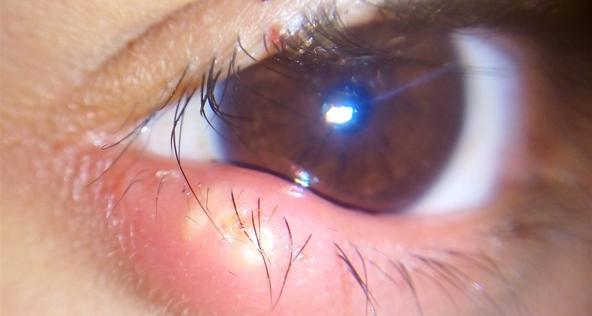
An eyelid bump that hurts is telling you something specific — and it usually is not the same thing as one that doesn’t.
A hordeolum, commonly called a stye, is an acute, painful infection of an eyelid gland.
It is one of the most common eyelid complaints seen in both primary care and ophthalmology.
Patients and clinicians frequently confuse it with a chalazion, but the two have different underlying processes and different urgency.
A hordeolum is infectious and acute; a chalazion is a sterile, chronic granulomatous reaction.
Getting that distinction right — along with knowing which gland is involved — is what actually guides appropriate treatment.
What Is a Hordeolum?
A hordeolum is an acute, typically staphylococcal infection of a gland at or near the eyelid margin.
It is classified by which gland is involved:
- External hordeolum — infection of a gland of Zeis or Moll at the base of an eyelash, presenting near the lid margin
- Internal hordeolum — infection of a meibomian gland within the tarsal plate, often presenting as a deeper, more diffuse swelling
Both forms are acute and tender, distinguishing them clinically from the painless, chronic swelling of a chalazion.
Epidemiology
Hordeola are extremely common and occur across all age groups.
- They are among the most frequent acute eyelid complaints presenting to primary care, urgent care, and eye clinics
- They can occur at any age, including children, and recurrence is common in patients with underlying lid margin disease
- External hordeola are generally more common than internal ones, given the larger number of superficial glands along the lash line
Because most cases resolve quickly with simple measures, many patients never seek formal medical care, so true incidence is likely underestimated.
All-fit Slit-Lamp Adapter
Record and share exactly what you see at the slit lamp. One adapter fits any slit lamp or surgical microscope — and any smartphone.
From Choroida — the team behind this sitePathophysiology
A hordeolum develops through acute bacterial infection and obstruction of the involved gland.
- Staphylococcus aureus is the most commonly implicated organism
- Bacteria infect and obstruct the gland duct, most often a gland of Zeis, Moll, or a meibomian gland
- Trapped secretions and bacterial proliferation drive acute local inflammation
- Neutrophilic infiltration produces the characteristic tenderness, erythema, and pointing seen on exam
- The lesion frequently localizes and drains spontaneously as it matures
This acute infectious process is what separates a hordeolum from a chalazion, which arises from sterile lipid extravasation rather than active infection.
Risk Factors
Ocular Risk Factors
- Chronic blepharitis and meibomian gland dysfunction
- Prior chalazion or hordeolum, since recurrence is common
- Contact lens wear with suboptimal hygiene
- Ocular rosacea
Systemic and Behavioral Risk Factors
- Poor eyelid or hand hygiene, including frequent eye rubbing or eye makeup use without adequate removal
- Diabetes mellitus, related to a general predisposition to skin and soft-tissue infection
- Childhood, given the frequency of lid margin colonization and hygiene-related exposure in this age group
Addressing underlying blepharitis is often what actually prevents the next episode, not just treating the current one.
Clinical Presentation
Symptoms
- Acute onset of eyelid pain, tenderness, and swelling, typically over hours to a few days
- Localized redness that worsens before it points and often begins to drain
- Mild tearing or foreign-body sensation from mechanical irritation
- Occasional mild preseptal swelling in more extensive cases, without true orbital involvement
Examination Findings
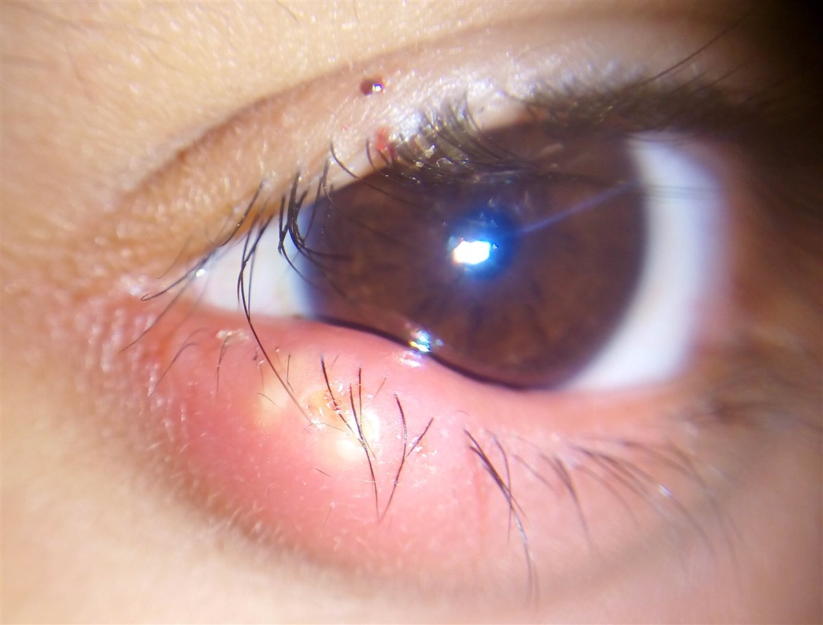
- A tender, erythematous, well-localized swelling at or near the lid margin
- A visible yellow-white point of pus in more mature external lesions
- External hordeola point toward the skin surface near the lash base; internal hordeola may point through the conjunctival surface
- Palpable warmth and tenderness on gentle palpation, unlike the painless nodule of a chalazion
- Mobile overlying skin, without the fixation or lash loss that would raise concern for a different eyelid lesion
Tenderness on palpation is the single most useful bedside sign separating an acute hordeolum from a chronic, painless chalazion.
Diagnostic Evaluation
Clinical Examination
- Diagnosis is clinical, based on the acute, tender, localized swelling at the lid margin
- Eyelid eversion helps localize an internal hordeolum and assess the degree of tarsal involvement
When Further Workup Is Warranted
- Spreading erythema, warmth, or swelling beyond the immediate lid margin — evaluate for preseptal cellulitis
- Proptosis, restricted eye movement, or pain with eye movement — evaluate urgently for orbital cellulitis
- Recurrent hordeola at the same site, or an atypical, non-resolving lesion — consider biopsy to exclude a masquerading lesion such as sebaceous gland carcinoma
Laboratory testing and imaging are rarely needed for a straightforward, localized hordeolum.
Differential Diagnosis
Conditions that can be confused with a hordeolum include:
- Chalazion — painless, chronic, non-tender nodule rather than an acute, tender, infected one
- Preseptal cellulitis — more diffuse eyelid erythema and swelling extending beyond a discrete point
- Dacryocystitis — swelling and tenderness centered over the lacrimal sac at the medial canthus rather than the lid margin
- Insect bite or contact dermatitis — history of exposure, often with itching rather than the acute pain of a hordeolum
- Sebaceous gland carcinoma — must be considered in a recurrent or non-resolving lesion, particularly in older patients
The combination of acute onset and tenderness is usually enough to separate a straightforward hordeolum from these mimics.
Management
Conservative Management
First-line therapy for essentially all uncomplicated cases:
- Warm compresses applied for 10-15 minutes, several times daily, to promote spontaneous drainage
- Gentle lid hygiene to reduce surrounding bacterial load and address any underlying blepharitis
- Avoiding eye makeup and contact lens wear until the lesion has resolved
Medical Therapy
- Topical antibiotic ointment (e.g., erythromycin) is commonly recommended, though evidence for benefit over compresses alone is limited
- Oral antibiotics are reserved for cases with significant surrounding erythema or when preseptal cellulitis is a concern
Incision and Drainage
- Reserved for large hordeola that fail to respond to warm compresses
- Performed with a fine-tipped blade once the lesion has clearly pointed, to avoid incising into non-purulent tissue
- Referral to ophthalmology is appropriate for large, refractory, or recurrent lesions
Most hordeola never need anything beyond warm compresses — escalation should be reserved for lesions that genuinely fail conservative care.
Prognosis
Prognosis is excellent for the overwhelming majority of cases.
- Most uncomplicated hordeola resolve within one to two weeks with warm compresses and lid hygiene
- Recurrence is common in patients with untreated underlying blepharitis or meibomian gland dysfunction
- Vision-threatening complications are rare and essentially limited to progression to preseptal or, very rarely, orbital cellulitis
Treating the underlying lid margin disease, not just the acute episode, is what actually reduces how often a patient comes back with another one.
Would you like to document anterior segment findings with your smartphone?


Document what you see
Two smartphone imaging tools built for everyday clinic use — one for the slit lamp, one for the fundus.
From Choroida — the team behind this siteReferences
- Hordeolum. StatPearls, NCBI Bookshelf.
- Chalazion and Hordeolum. Merck Manual, Professional Edition.
- Lindsley K, Nichols JJ, Dickersin K. Interventions for acute internal hordeolum. Cochrane Database of Systematic Reviews. 2017.
- American Optometric Association. Hordeolum (Stye).
- Hordeolum. EyeWiki, American Academy of Ophthalmology.
Test yourself
A few questions straight from this article.
-
A hordeolum (stye) is best described as which type of eyelid lesion?
A hordeolum is an acute, typically staphylococcal infection of a gland at or near the eyelid margin, which is why it is painful and tender rather than quietly chronic. -
Which gland is infected in an internal hordeolum?
Internal hordeola involve a meibomian gland inside the tarsal plate and therefore present as deeper, more diffuse swelling, sometimes pointing through the conjunctival surface. -
Which organism is most commonly implicated in a hordeolum?
Staphylococcus aureus is the organism most often responsible; it infects and obstructs the gland duct, and trapped secretions with bacterial proliferation drive the acute inflammation. -
A chalazion arises from which process, unlike the acute infection of a hordeolum?
A chalazion is a sterile, chronic granulomatous reaction to extravasated lipid, whereas a hordeolum is an active bacterial infection of the gland. -
Which bedside sign best separates an acute hordeolum from a chalazion?
Tenderness on gentle palpation is the single most useful bedside sign, since a chalazion presents as a painless nodule while a hordeolum is acutely tender and warm. -
What is first-line treatment for an uncomplicated hordeolum?
Warm compresses for 10-15 minutes several times daily, with gentle lid hygiene, promote spontaneous drainage and are first-line for essentially all uncomplicated cases. -
Within what period do most uncomplicated hordeola resolve with conservative care?
Most uncomplicated hordeola settle within one to two weeks using warm compresses and lid hygiene, and the overall prognosis is excellent. -
Which finding in a patient with a hordeolum prompts urgent evaluation for orbital cellulitis?
Proptosis, restricted eye movement, or pain on eye movement signals possible orbital involvement and warrants urgent evaluation rather than continued conservative care. -
A recurrent, non-resolving eyelid lesion in an older patient should prompt biopsy to exclude what?
Recurrent hordeola at the same site or an atypical, non-resolving lesion raise concern for a masquerading malignancy, particularly sebaceous gland carcinoma in older patients. -
Which measure most reduces how often a patient returns with another hordeolum?
Recurrence is common when lid margin disease goes untreated, so addressing underlying blepharitis and meibomian gland dysfunction does more to prevent the next episode than treating the current one alone.
