Pinguecula vs pterygium is a distinction that matters for follow-up and surgery.
Both are fibrovascular degenerations of the bulbar conjunctiva.
Only the pterygium grows onto the cornea.
What Is a Pinguecula
A pinguecula is a yellowish raised patch next to the limbus.
It lies on the nasal or temporal conjunctiva in the palpebral fissure.
It does not cross onto the cornea (see pinguecula).
Most are painless and stable.
What Is a Pterygium
A pterygium is a triangular wing of tissue that crosses the limbus.
It extends onto the cornea and can reach the pupil.
It is more often nasal.
Therefore, it can cause blur and astigmatism.
All-fit Slit-Lamp Adapter
Record and share exactly what you see at the slit lamp. One adapter fits any slit lamp or surgical microscope — and any smartphone.
From Choroida — the team behind this siteShared Causes
Ultraviolet exposure, wind and dust drive both conditions.
Outdoor workers and people in sunny climates have higher rates.
Dry eye adds to the risk.
So advice on protection applies to both.
Pinguecula vs Pterygium on Examination
Look at the limbus with a slit lamp.
A pinguecula stops at the limbus.
A pterygium has a head on the cornea and a body on the conjunctiva.
Also look for a Stocker line, which is an iron line at the head.
Symptoms
A pinguecula causes irritation, dryness and cosmetic concern.
A pterygium also causes redness and foreign body sensation.
It may reduce vision as it advances.
Therefore, check acuity and topography when it is large.
Inflamed Lesions
Both lesions can become red and tender, a state called pingueculitis.
Lubricants and a short course of a mild steroid help.
However, repeated flares should prompt a review of dryness.
So treat the surface as well as the lesion.
When Observation Is Enough
Most pingueculae need no treatment.
Small, stable pterygia also need only monitoring.
Document size with photographs.
Also review annually.
Treatment of Pterygium
Surgery is considered for visual loss, astigmatism, motility restriction or persistent inflammation.
Conjunctival autograft lowers the risk of recurrence (see conjunctival autograft).
Recurrence is the main concern (see pterygium recurrence).
So choose the technique carefully.
Pinguecula vs Pterygium and Cosmetic Surgery
Some patients request removal for appearance.
Counsel them on recurrence and scarring.
Also discuss that surgery does not remove the cause.
Therefore, individualize the decision.
Protecting the Eyes
Sunglasses with ultraviolet protection and a wide-brim hat help.
Lubricants may reduce irritation.
Also avoid dusty environments when possible.
As a result, progression may slow.
Pitfalls to Avoid
- Do not call a lesion a pinguecula without checking the limbus
- Never operate without documenting astigmatism
- Avoid long steroid courses for recurrent redness
- Do not forget biopsy for atypical lesions
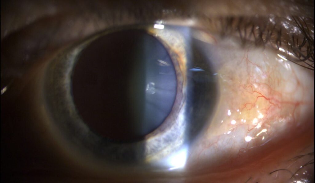
Pinguecula vs Pterygium on Imaging
Slit-lamp photographs record the extent of both lesions.
Anterior segment OCT shows the depth of invasion.
Topography documents induced astigmatism.
Therefore, imaging helps decide when to operate.
Pinguecula vs Pterygium and Astigmatism
A pterygium pulls on the cornea and induces with-the-rule astigmatism.
Larger lesions cause greater distortion.
After removal, astigmatism often falls.
So measure it before and after surgery.
Pinguecula vs Pterygium and Contact Lenses
Raised lesions disturb lens fit and comfort.
Lenses may rub the head of a pterygium.
Switch to a smaller lens or a different design.
Also treat dryness to improve comfort.
Pinguecula vs Pterygium in Young Patients
Young patients with rapid growth need prompt review.
Such lesions are more likely to recur after surgery.
Counsel them on sun protection from the start.
Also treat inflammation early.
Pinguecula vs Pterygium: Red Flags
Rapid growth, pigment or a feeding vessel raise the concern for a tumor.
Ocular surface squamous neoplasia can mimic a pterygium.
Biopsy any lesion that looks unusual.
In addition, send excised tissue for pathology.
Pinguecula vs Pterygium: Patient Questions
Patients ask whether the lesion will turn into cancer.
Most do not, but monitoring is wise.
They also ask if drops can remove it.
Drops relieve inflammation but do not remove the tissue.
Pinguecula vs Pterygium and Lifestyle
Wear sunglasses outdoors.
Use lubricants in dry or windy weather.
Also avoid smoking, which adds irritation.
Therefore, small changes help over years.
Pinguecula vs Pterygium Follow-Up
Review stable lesions yearly.
Review growing lesions more often.
Record size with the same method each time.
This makes progression clear.
Pinguecula vs Pterygium and Dry Eye
Both lesions disturb the tear film.
Patients feel dryness and burning over the lesion.
Treat the surface to ease symptoms.
Lubricants and lid care are the first steps.
Pinguecula vs Pterygium: When to Operate
Operate for visual loss, motility limits or repeated inflammation.
Consider cosmetic requests after discussion.
Choose the technique with care to lower recurrence.
Therefore, document the reason for surgery.
Practical Summary
If the lesion stops at the limbus, call it a pinguecula.
When it crosses onto the cornea, call it a pterygium.
Because progression is slow, monitoring is usually enough.
However, operate when vision or comfort suffers.
For the latest studies, see the PubMed results for pinguecula vs pterygium.


Document what you see
Two smartphone imaging tools built for everyday clinic use — one for the slit lamp, one for the fundus.
From Choroida — the team behind this siteReferences
- Chu WK, Choi HL, Bhat AK, Jhanji V. Pterygium: new insights. Eye. 2020;34:1047-1050.
- Coroneo MT, Di Girolamo N, Wakefield D. The pathogenesis of pterygia. Curr Opin Ophthalmol. 1999;10:282-288.
- Hirst LW. The treatment of pterygium. Surv Ophthalmol. 2003;48:145-180.
- Shields JA, Shields CL. Eyelid, Conjunctival, and Orbital Tumors: An Atlas and Textbook. 3rd ed. Wolters Kluwer; 2016.

Writes and reviews the clinical articles on Educate. Every post is checked against peer-reviewed sources before it goes live.