
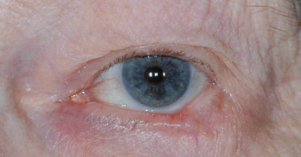
Periocular basal cell carcinoma is the most common malignant eyelid tumor by a wide margin, and although it rarely metastasizes, it can invade locally into the orbit, nasolacrimal system, or sinuses if left untreated, particularly when it arises at the medial canthus.
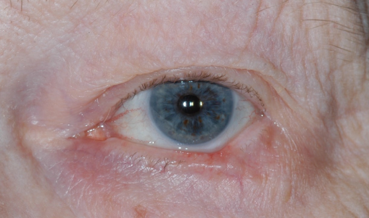
Cure rates are excellent with appropriate excision, so the greater challenge is usually reconstruction, which has to be planned around the specific site involved rather than following a single formula for every eyelid defect.
Clinical Presentation
Basal cell carcinoma most often affects the lower eyelid, followed by the medial canthus, upper eyelid, and lateral canthus in decreasing frequency.
- Nodular type: a pearly, telangiectatic nodule, sometimes with central ulceration, the most common and most recognizable pattern
- Ulcerative type: a rodent ulcer with a rolled, pearly border and central crater
- Morpheaform (sclerosing) type: a flat, indurated, scar-like plaque with indistinct margins, harder to detect clinically and prone to deeper, more extensive subclinical spread
- Superficial type: a scaly, erythematous patch, less common on the eyelid than elsewhere on the skin
Morpheaform tumors are the most likely to be under-recognized clinically and to extend well beyond their apparent visible margin.
Why the Medial Canthus Matters
Tumors at the medial canthus have direct access to the orbit, nasolacrimal drainage system, and ethmoid sinuses through relatively unobstructed tissue planes, so periocular basal cell carcinoma at this site carries a higher risk of deep invasion than tumors elsewhere on the eyelid.
Any medial canthal lesion suspicious for basal cell carcinoma deserves prompt biopsy, and confirmed tumors at this site need particularly careful margin assessment before reconstruction is planned.
All-fit Slit-Lamp Adapter
Record and share exactly what you see at the slit lamp. One adapter fits any slit lamp or surgical microscope — and any smartphone.
From Choroida — the team behind this siteDiagnosis and Excision
Biopsy confirms the diagnosis and histologic subtype before definitive treatment.
Mohs micrographic surgery is preferred for periocular basal cell carcinoma because it confirms complete margin clearance while sparing as much normal eyelid tissue as possible, which matters more here than almost anywhere else on the body given how little spare tissue the eyelid has.
Morpheaform and recurrent tumors, and those at the medial canthus, particularly benefit from Mohs surgery given their tendency toward deeper or wider subclinical extension.
Reconstruction by Site
Lower Eyelid
Small to moderate defects can often be closed directly or with a lateral canthotomy and cantholysis for added laxity; larger full-thickness defects typically need a tarsoconjunctival flap for the posterior lamella combined with an anterior lamellar skin graft or flap (see eyelid margin tumor reconstruction).
Medial Canthus
Medial canthal defects are reconstructed with attention to the lacrimal drainage system, which may need to be repaired or bypassed if the canaliculi are involved, and local flaps such as a glabellar or rhomboid flap are commonly used to restore the concave canthal contour.
Upper Eyelid
Upper eyelid defects are reconstructed conservatively because of the greater functional consequence of tightness or malposition here, prioritizing full closure and corneal protection over aggressive cosmetic tension.
Lateral Canthus
Lateral canthal defects often use a periosteal flap or lateral tarsal strip technique to re-anchor the eyelid to the orbital rim.
Follow-Up
Patients with a history of periocular basal cell carcinoma remain at risk for new primary tumors elsewhere on sun-exposed skin, so ongoing dermatologic surveillance is appropriate.
Recurrence after Mohs surgery is uncommon but is checked for at follow-up visits, along with assessment of eyelid function, tear film, and corneal protection after reconstruction.
Prognosis
Cure rates after Mohs excision are very high, and metastasis is rare.
The functional and cosmetic outcome depends more on the reconstruction than on the tumor itself in most cases, which is why matching the repair to the specific eyelid site is as important as achieving clear margins.


Document what you see
Two smartphone imaging tools built for everyday clinic use — one for the slit lamp, one for the fundus.
From Choroida — the team behind this siteReferences
- Malhotra R, Huilgol SC, Huynh NT, Selva D. The Australian Mohs database, part I: periocular basal cell carcinoma experience over 7 years. Ophthalmology. 2004;111:624-630.
- Cook BE Jr, Bartley GB. Treatment options and future prospects for the management of eyelid malignancies: an evidence-based update. Ophthalmology. 2001;108:2088-2098.
- Leibovitch I, McNab A, Sullivan T, Davis G, Selva D. Orbital invasion by periocular basal cell carcinoma. Ophthalmology. 2005;112:717-723.
- Rathi VM, Sudharshan S, Biswas J. Eyelid basal cell carcinoma: outcome of excision and reconstruction. Indian J Ophthalmol. 2004;52:337-338.
Test yourself
A few questions straight from this article.
-
Which eyelid site is most frequently involved by periocular basal cell carcinoma?
Basal cell carcinoma most often affects the lower eyelid, followed in decreasing frequency by the medial canthus, upper eyelid and lateral canthus. -
What is the characteristic biological behaviour of periocular basal cell carcinoma?
It rarely metastasizes, but untreated tumours invade locally into the orbit, the nasolacrimal system or the sinuses, particularly from the medial canthus. -
Which description matches the nodular form of periocular basal cell carcinoma?
The nodular pattern is the most common and most recognizable: a pearly, telangiectatic nodule that sometimes shows central ulceration. -
Which subtype of periocular basal cell carcinoma extends furthest beyond its visible margin?
The morpheaform or sclerosing type is a flat, indurated plaque with indistinct margins; it is the most often under-recognized clinically and spreads furthest subclinically. -
Why does medial canthal basal cell carcinoma carry a higher risk of deep invasion?
Medial canthal tumours have direct access to the orbit, the nasolacrimal drainage system and the ethmoid sinuses through relatively unobstructed tissue planes. -
Why is Mohs micrographic surgery preferred for periocular basal cell carcinoma?
Mohs confirms complete margin clearance while preserving as much normal eyelid tissue as possible, which matters more on the eyelid than almost anywhere else. -
A large full-thickness lower eyelid defect is typically repaired with which combination?
Larger full-thickness lower lid defects usually need a tarsoconjunctival flap for the posterior lamella combined with an anterior lamellar skin graft or flap. -
Which local flap commonly restores the concave contour of the medial canthus?
Medial canthal defects are repaired with attention to lacrimal drainage, and local flaps such as a glabellar or rhomboid flap are used to rebuild the concave canthal contour. -
Upper eyelid reconstruction after tumour excision prioritizes which goal?
Because tightness or malposition has greater functional consequence in the upper lid, repair is conservative and prioritizes complete closure and corneal protection. -
Why is long-term dermatologic surveillance advised after periocular basal cell carcinoma?
These patients stay at risk of new primary tumours elsewhere on sun-exposed skin, so dermatologic surveillance continues alongside checks of eyelid function and corneal protection.
