
DISEASE “Xanthelasma”
Xanthelasma Palpebrarum (XP) is a benign condition involving cutaneous lesions in the periocular region. It presents as a xanthoma found on the medial aspect of the eyelids.
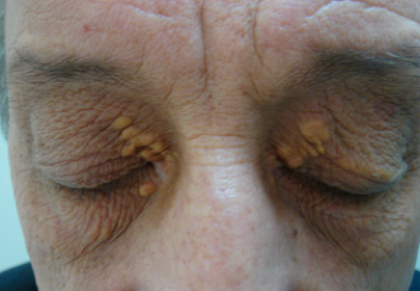
These lesions do not typically present with functional limitations. It occurs more commonly in women (1.2% incidence rate) than in men (0.3% incidence rate) with a peak incidence at the ages of 30-50 years old.
Nearly half of patients with XP have associated lipid disorders. These include Type II hyperlipidemia, Type IV hyperlipidemia, high total cholesterol, high triglycerides, and low levels of high-density lipoprotein (HDL).
However, despite the strong association of XP with lipid imbalance, 25-70% of patients are normolipidemic on presentation.
Xanthelasma Diagnosis
XP lesions present as soft, yellow papules or plaques filled with cholesterol at the medial canthus of the eyelids. They can appear as multiple, symmetric lesions that more commonly are found on the upper eyelid than the lower one.

Patients will seek professional treatment for xanthelasma due to cosmetic concerns.
Additionally, these lesions tend to be permanent and can grow in size with time which may be distressing. From a provider standpoint, the diagnosis can be made clinically.
Biopsy:
Reticular dermis infiltration of foamy macrophages with sparing of the epidermis, papillary dermis, and subcutaneous fatty layer.
The inflammatory infiltrates in the superficial reticular dermis will be perivascular and consist of foam cells with lipid-laden cytoplasmic vacuoles.
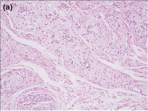
Foam cells, or xanthoma cells, originate from macrophages that have accumulated intracellular cholesterol, lipids, and fat.
All-fit Slit-Lamp Adapter
Record and share exactly what you see at the slit lamp. One adapter fits any slit lamp or surgical microscope — and any smartphone.
From Choroida — the team behind this siteMANAGEMENT of Xanthelasma
Conservative therapy begins with a low-fat diet and statins- the therapeutic effect is questionable. Popular treatment options include surgical excision, laser therapy, chemical peel, cryotherapy, and radiofrequency ablation.
Surgical excision of the xanthelasma was one of the earliest treatments, however, post-operative adverse outcomes must be considered.
Risk of cicatricial ectropion, transient hematomas, poor cosmetic outcome, recurrence, and infection are possible.
Trichloroacetic acid (TCA) is commonly used for the peel at a 70% concentration.
Laser therapy utilizes energy to coagulate vessels and damage the perivascular foam cells that make up the lesion.
Cryotherapy causes vasoconstriction and the formation of microthrombi as a result of cell death within XP lesions.
Radiofrequency treatment works by causing electrocoagulation for instant hemostasis and is used in vascular lesions.


Document what you see
Two smartphone imaging tools built for everyday clinic use — one for the slit lamp, one for the fundus.
From Choroida — the team behind this siteREFERENCES
1. Esmat SM, Elramly AZ, Abdel Halim DM, Gawdat HI, Taha HI. Fractional CO2 laser is an effective therapeutic modality for xanthelasma palpebrarum: a randomized clinical trial. Dermatol Surg. Dec 2014;40(12):1349-55. doi:10.1097/DSS.0000000000000172
2. Aggarwal R, Rathore PK. A study evaluating xanthelasma palpebrarum clinically and biochemically. International Journal of Contemporary Medical Research. 2016;3(9):2565-7.
3. Dincer D, Koc E, Erbil AH, Kose O. Effectiveness of low-voltage radiofrequency in the treatment of xanthelasma palpebrarum: a pilot study of 15 cases. Dermatol Surg. Dec 2010;36(12):1973-8. doi:10.1111/j.1524-4725.2010.01770.x
4. Nahas TR, Marques JC, Nicoletti A, Cunha M, Nishiwaki-Dantas MC, Filho JV. Treatment of eyelid xanthelasma with 70% trichloroacetic acid. Ophthalmic Plast Reconstr Surg. Jul-Aug 2009;25(4):280-3. doi:10.1097/IOP.0b013e3181aa9a1f
5. Al Aboud AM, Al Aboud DM. Xanthelasma Palpebrarum. StatPearls. 2021.
Test yourself
A few questions straight from this article.
-
Where on the eyelids does xanthelasma palpebrarum characteristically appear?
Xanthelasma palpebrarum is a benign periocular cutaneous xanthoma found on the medial aspect of the eyelids, and it does not usually limit lid function. -
Which pattern of occurrence is reported for xanthelasma palpebrarum?
The incidence is 1.2% in women against 0.3% in men, with the peak occurring between the ages of 30 and 50 years. -
Roughly what proportion of patients with xanthelasma palpebrarum have an associated lipid disorder?
Nearly half of patients have an associated lipid disorder, including type II or type IV hyperlipidaemia, raised total cholesterol or triglycerides, and low HDL. -
What proportion of patients with xanthelasma palpebrarum are normolipidemic when they present?
Despite the strong association with lipid imbalance, between 25% and 70% of patients have normal lipids at presentation, so normal results do not exclude the diagnosis. -
How do xanthelasma lesions typically appear on examination?
They present as soft, yellow, cholesterol-filled papules or plaques at the medial canthus, often multiple and symmetric, and more often on the upper lid than the lower. -
How is the diagnosis of xanthelasma palpebrarum usually established?
From the provider's standpoint the diagnosis can be made clinically; biopsy is not required to recognise the lesion. -
What does biopsy of a xanthelasma lesion show?
Biopsy shows reticular dermis infiltration by foamy macrophages, with sparing of the epidermis, papillary dermis and subcutaneous fatty layer; the infiltrate is perivascular and made of lipid-laden foam cells. -
What does the article say about conservative therapy with a low-fat diet and statins in xanthelasma?
Conservative therapy begins with a low-fat diet and statins, but the article states that the therapeutic effect of this approach is questionable. -
Which adverse outcome must be considered after surgical excision of xanthelasma?
Surgical excision carries a risk of cicatricial ectropion, along with transient haematoma, poor cosmetic outcome, recurrence and infection. -
At what concentration is trichloroacetic acid commonly used for chemical peeling of xanthelasma?
Trichloroacetic acid is commonly used for the peel at a 70% concentration, one of several destructive options alongside laser, cryotherapy and radiofrequency.
