DME OCT patterns describe how fluid distributes in the macula of an eye with diabetic macular edema.
Three classic patterns are diffuse thickening, cystoid edema and serous detachment.
Therefore, reading the pattern helps you judge the mechanism and plan treatment.
Why Describe DME OCT Patterns
Central thickness is a single number and hides the shape of the disease.
Patterns show where the fluid sits and what else is present.
Also, they help compare eyes and follow changes.
Because treatment differs by pattern, the description has practical value.
The Classic Description of DME OCT Patterns
Otani and colleagues described three patterns in 1999.
Sponge-like diffuse thickening, cystoid macular edema and serous retinal detachment became the basic classification.
Also, many eyes show a mix of patterns.
Therefore, treat the classification as a framework.
Fundus Explorer Pro
Photograph the retinal findings described here with the phone already in your pocket — 22 D optics and built-in illumination in one handheld unit.
From Choroida — the team behind this siteDiffuse Retinal Thickening
The retina looks thick and spongy with reduced reflectivity.
Small cystic spaces may be present but are not prominent.
Also, diffuse leakage from capillaries and microaneurysms is the usual cause.
Because the disease is widespread, laser alone has limited effect.
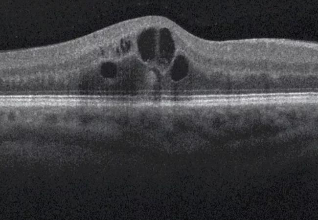
Cystoid Macular Edema
Large cystic spaces form in the outer plexiform layer and the inner nuclear layer.
They may coalesce into a large central cavity.
Also, long-standing cystoid edema is linked with damage to the outer retina (see cystoid macular edema).
Therefore, treat cystoid edema promptly.
Serous Retinal Detachment
A pocket of subretinal fluid lies under the fovea.
Gaucher and colleagues described its evolution on OCT.
Also, some studies suggest that eyes with subretinal fluid respond well to anti-VEGF drugs.
Because the fluid indicates breakdown of the outer barrier, it deserves attention.
Tractional DME OCT Patterns
Kim and colleagues added patterns with vitreomacular traction and epiretinal membranes.
These eyes may respond less to injections (see vitreomacular traction).
Also, vitrectomy may help in selected cases.
Therefore, look at the vitreoretinal interface.
Hard Exudates
Hyperreflective deposits in the outer retina mark lipid exudates.
They accompany long-standing leakage.
Also, they can be a cause of poor vision when they enter the fovea (see hard exudates).
Because they respond slowly, patients need time.
Outer Retinal Integrity
The ellipsoid zone and the external limiting membrane predict vision.
Disruption indicates photoreceptor loss (see ellipsoid zone disruption).
Also, disorganization of the inner layers carries a poor prognosis.
Therefore, look beyond the fluid.
DME OCT Patterns and Treatment Choice
Anti-VEGF therapy is the first choice for center-involving edema.
Steroids help in some resistant eyes (see steroids in DME).
Also, focal laser treats leaking microaneurysms in selected cases.
Because the pattern suggests the mechanism, it guides the order of options.
Following Response
Measure central thickness and look at the pattern at each visit.
A pattern that changes from diffuse to cystoid may signal recurrence.
Also, residual fluid can persist despite good vision.
Therefore, combine imaging with acuity.
Limits of DME OCT Patterns
Patterns overlap and observers disagree.
Image quality and segmentation also affect interpretation.
Also, no pattern replaces clinical judgment.
Documenting DME OCT Patterns
Record the type, the central thickness and the presence of traction or exudate.
Also, save the scan for comparison.
OCT Biomarkers Beyond Fluid
Hyperreflective foci are small dots in the retina and may reflect inflammatory cells or lipid.
Also, disorganization of the inner retinal layers is a negative sign.
Because these markers predict outcome, report them.
Response to Anti-VEGF by Pattern
Eyes with subretinal fluid often respond well.
Eyes with cystoid change and exudates respond more slowly.
Also, eyes with traction respond poorly.
Therefore, consider alternatives when the response is poor.
Comparing Eyes Over Time
Use the same device and the same scan protocol.
Align the scans with the same landmarks.
Also, record changes in central thickness.
Because device differences matter, avoid mixing machines.
Cystoid Space Size and Visual Acuity
Large cysts may be associated with worse acuity.
Also, cysts in the outer layers carry more risk for photoreceptor damage.
Therefore, combine the pattern with acuity.
Teaching Points
Describe the findings in simple words.
Show the fluid on the scan to the patient.
Also, explain that injections aim to dry the retina.
Because patients adhere better when they understand, use the images.
Pitfalls
Do not confuse cystoid spaces with outer retinal tubulation.
Also, do not overlook the vitreoretinal interface.
Therefore, scan the whole macula.


Document what you see
Two smartphone imaging tools built for everyday clinic use — one for the slit lamp, one for the fundus.
From Choroida — the team behind this siteReferences
- Otani T, Kishi S, Maruyama Y. Patterns of diabetic macular edema with optical coherence tomography. Am J Ophthalmol. 1999;127:688-693.
- Panozzo G, Parolini B, Gusson E, et al. Diabetic macular edema: an OCT-based classification. Semin Ophthalmol. 2004;19:13-20.
- Kim BY, Smith SD, Kaiser PK. Optical coherence tomographic patterns of diabetic macular edema. Am J Ophthalmol. 2006;142:405-412.
- Gaucher D, Sebah C, Erginay A, et al. Optical coherence tomography features during the evolution of serous retinal detachment in patients with diabetic macular edema. Am J Ophthalmol. 2008;145:289-296.

Writes and reviews the clinical articles on Educate. Every post is checked against peer-reviewed sources before it goes live.