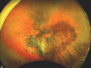
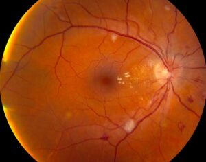
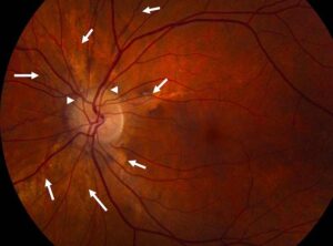
Retinoblastoma classification sorts each affected eye according to the extent of intraocular disease.
The International Classification of Retinoblastoma (ICRB) runs from group A to group E.
Generally, later groups carry a lower chance of saving the eye.
Therefore, the group helps clinicians plan treatment and counsel families (see retinoblastoma overview).
Why Retinoblastoma Classification Changed
Reese and Ellsworth published their grouping in 1963, when external beam radiotherapy was central to globe-sparing care.
Later, chemotherapy replaced radiotherapy as the main approach in many centers.
Therefore, clinicians needed a system that matched modern treatment.
Murphree proposed a new group system in 2005, with greater emphasis on seeds and subretinal fluid.
Examining for Retinoblastoma Classification
Clinicians usually assign the group during an examination under anesthesia.
Indirect ophthalmoscopy with scleral depression shows tumor location, seeds and retinal detachment.
Next, wide-field photographs document the baseline, while ultrasound and MRI add detail when needed.
Fundus Explorer Pro
Photograph the retinal findings described here with the phone already in your pocket — 22 D optics and built-in illumination in one handheld unit.
From Choroida — the team behind this siteGroup A: Small Tumors Away From the Fovea
Group A tumors measure 3 mm or less and stay within the retina.
They also lie at least 3 mm from the foveola and 1.5 mm from the optic disc.
Focal laser or cryotherapy may control these lesions.
However, children with germline disease still need close surveillance for new tumors.
Group B: Larger Tumors Without Seeds
Group B includes other retina-confined tumors without vitreous or subretinal seeds.
For example, a small macular tumor falls here because of its location.
Treatment usually combines chemotherapy with focal consolidation.
Even so, a salvaged eye with a macular tumor may keep poor central vision.
Group C: Focal Seeds
Group C describes localized seeding within 3 mm of a tumor.
The seeds may lie in the vitreous, the subretinal space or both.
Because disease remains focal, globe salvage is often realistic.
Group D: Diffuse Seeds
Group D eyes show seeding more than 3 mm from the tumor.
Extensive subretinal fluid or total retinal detachment may also be present.
Consequently, the prognosis for salvage becomes less predictable.
Teams may consider intra-arterial or intravitreal chemotherapy (see retinoblastoma treatment options).
Primary enucleation also remains reasonable, particularly in unilateral disease.
Group E: Eyes With Extensive Disease
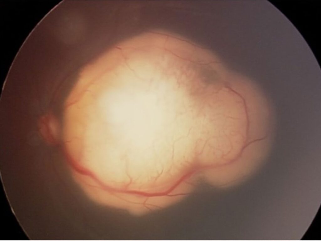
Group E eyes have advanced disease and usually little visual potential.
Suspect this group when the examination shows any of these features:
- Tumor touching the lens
- Neovascular glaucoma
- Anterior segment or ciliary body involvement
- Diffuse infiltrating retinoblastoma
- Media opacity from hemorrhage
Two Versions of Retinoblastoma Classification
Published ICRB versions differ, and this creates confusion.
The Philadelphia version by Shields defines group E partly by tumor filling over half the globe.
Therefore, reports should state which version they used.
Otherwise, results from different centers may not compare fairly.
Retinoblastoma Classification and Chemotherapy Outcomes
Shields and colleagues tested the ICRB in 249 eyes treated with chemoreduction.
Treatment success reached 100% in group A, 93% in group B, 90% in group C and 47% in group D.
These figures came from one center, so they cannot predict an individual outcome.
Additionally, they do not transfer directly to intra-arterial protocols.
How Seed Type Refines the Group
Munier described vitreous seeds as dust, spheres or clouds.
However, ICRB grouping does not capture this morphology, so clinicians should record it separately.
Retinoblastoma Classification and Enucleation Decisions
Group alone should never dictate enucleation.
Clinicians also weigh laterality, visual potential, germline status and the fellow eye.
Meanwhile, a blind unilateral group E eye usually favors primary enucleation.
Pathology then looks for high-risk features, including massive choroidal or postlaminar optic nerve invasion.
What Retinoblastoma Classification Cannot Tell You
The ICRB describes intraocular disease at presentation.
It cannot confirm metastatic spread or replace histopathology after enucleation.
It also cannot establish germline status, so families still need genetic counseling.
Moreover, mimics such as ocular toxocariasis can resemble tumor.
Counseling Families About Retinoblastoma Classification
Start by explaining that the group describes the eye, and survival depends on other factors.
Next, separate three goals clearly: saving life, saving the eye and saving vision.
Families often assume that a salvaged globe will see well.
However, tumor location, detachment and treatment complications may still limit vision.
Pitfalls to Avoid
- Do not assign a group without a complete dilated examination
- Avoid fine needle biopsy when retinoblastoma remains possible
- Do not mix ICRB versions when comparing outcomes
- Never regroup an eye after treatment response
- Do not equate globe salvage with useful vision


Document what you see
Two smartphone imaging tools built for everyday clinic use — one for the slit lamp, one for the fundus.
From Choroida — the team behind this siteReferences
- Murphree AL. Intraocular retinoblastoma: the case for a new group classification. Ophthalmol Clin North Am. 2005;18:41-53.
- Shields CL, Mashayekhi A, Au AK, et al. The International Classification of Retinoblastoma predicts chemoreduction success. Ophthalmology. 2006;113:2276-2280.
- Reese AB, Ellsworth RM. The evaluation and current concept of retinoblastoma therapy. Trans Am Acad Ophthalmol Otolaryngol. 1963;67:164-172.
- Munier FL. Classification and management of seeds in retinoblastoma. Ophthalmic Genet. 2014;35:193-207.
- Shields CL, Manjandavida FP, Lally SE, et al. Intra-arterial chemotherapy for retinoblastoma in 70 eyes: outcomes based on the International Classification of Retinoblastoma. Ophthalmology. 2014;121:1453-1460.

Writes and reviews the clinical articles on Educate. Every post is checked against peer-reviewed sources before it goes live.