
Corneal iron lines all share the same underlying mechanism, iron from the tear film depositing within the corneal epithelium at a site where normal tear flow and blinking dynamics have been locally disrupted, and recognizing which specific named line is present, based on its precise location, can itself point directly to the underlying anatomic or pathologic cause responsible for that local disruption.
The Shared Mechanism
Iron is a normal trace component of the tear film, and under ordinary circumstances it is distributed and cleared evenly across the corneal surface with each blink.
When some local factor disrupts this normal, even tear film distribution, whether a change in corneal curvature, an adjacent surface lesion, or an altered pattern of eyelid closure, iron can accumulate and deposit within the corneal epithelium specifically at that site, over time becoming visible as a line or band on slit-lamp examination, best seen with a cobalt blue filter or, in some cases, on careful examination with white light alone.
Hudson-Stahli Line
Located horizontally at the junction of the upper two-thirds and lower one-third of the cornea, roughly corresponding to the lower eyelid margin position, this is the most commonly encountered corneal iron line, an age-related finding thought to relate to the normal pattern of tear film pooling and blink dynamics at this specific horizontal position, generally considered an incidental finding without independent clinical significance.
All-fit Slit-Lamp Adapter
Record and share exactly what you see at the slit lamp. One adapter fits any slit lamp or surgical microscope — and any smartphone.
From Choroida — the team behind this siteFleischer Ring
A ring or partial ring of iron deposition at the base of the cone in keratoconus, resulting from the altered tear film dynamics over the ectatic, abnormally shaped cornea, and considered one of the classic supportive slit-lamp signs of this condition (see keratoconus slit-lamp signs).
Stocker Line
A vertical or curved iron line found at the leading (advancing) edge of a pterygium, resulting from the local alteration in tear film flow created by the pterygium’s elevated, encroaching tissue, and its presence can sometimes provide a subtle clue to the direction and pattern of pterygium growth.
Ferry Line
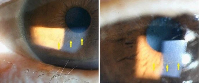
An iron line found adjacent to a filtering bleb following glaucoma filtering surgery, resulting from the altered local tear film dynamics created by the bleb’s presence on the ocular surface.
Other Recognized Iron Lines
- A line can form adjacent to areas of altered corneal curvature following refractive surgery, related to the same tear-flow-disruption mechanism operating around any change in corneal surface contour
- Iron lines have also been described in association with other conditions that locally alter corneal shape or the overlying tear film pattern, reinforcing that this is a general phenomenon occurring wherever local tear dynamics are sufficiently disrupted, not a finding unique to any single condition
Clinical Significance
Corneal iron lines are, in themselves, entirely benign and asymptomatic, never threatening vision or requiring any treatment directed at the iron deposition itself.
Their real clinical value lies in what they indicate about the underlying corneal surface or adjacent lesion: a Hudson-Stahli line is simply reassuring, expected background information, while a Fleischer ring supports a keratoconus diagnosis, a Stocker line confirms and characterizes an adjacent pterygium, and a Ferry line confirms the presence and local effect of a filtering bleb.
Examination Technique
Cobalt blue illumination at the slit lamp enhances the visibility of these iron deposits considerably compared with white light alone, since iron produces a distinctive appearance under this specific illumination, making cobalt blue examination a valuable addition whenever an iron line is suspected or being specifically sought as a supportive sign for another diagnosis.
Management
No treatment is directed at corneal iron lines themselves, since they are asymptomatic and clinically insignificant in their own right; management, when relevant, is entirely directed at the underlying condition the specific line has helped to identify or characterize, whether that is keratoconus, a pterygium, or a filtering bleb.
Clinical Value
Recognizing and correctly naming the specific corneal iron line present during a slit-lamp examination is a small but genuinely useful piece of diagnostic pattern recognition, turning what might otherwise be dismissed as an incidental pigmented line into a specific, informative clue about the local corneal surface anatomy and any adjacent pathology driving its formation.


Document what you see
Two smartphone imaging tools built for everyday clinic use — one for the slit lamp, one for the fundus.
From Choroida — the team behind this siteReferences
- Norn MS. Hudson-Stahli’s line of the cornea. IV. Follow-up examination. Acta Ophthalmol (Copenh). 1968;46:679-685.
- Sturrock GD. Iron lines in the anterior cornea. Trans Ophthalmol Soc U K. 1976;96:437-442.
- Rabinowitz YS. Keratoconus. Surv Ophthalmol. 1998;42:297-319.
- Ferry AP. A “new” iron line of the superficial cornea. Occurrence in patients with filtering blebs. Arch Ophthalmol. 1968;79:142-145.

Writes and reviews the clinical articles on Educate. Every post is checked against peer-reviewed sources before it goes live.


