CRAO recovery is usually limited, and most eyes keep severe central vision loss after a complete occlusion.
However, outcomes vary because residual retinal flow differs between patients.
Therefore, the first examination offers useful clues about what vision may return.
What CRAO Recovery Usually Looks Like
Many patients present with counting fingers vision or worse.
Without treatment, meaningful improvement occurs in a minority of these eyes.
For example, one patient-level meta-analysis reported recovery in about 18% of conservatively managed patients.
However, studies define recovery differently, so quote any single figure cautiously.
Why the Inner Retina Recovers Poorly
The central retinal artery supplies the inner retina, including its ganglion cells.
These neurons tolerate ischemia poorly and do not regenerate.
In primate work, Hayreh found no detectable damage after about 97 minutes of occlusion.
By 240 minutes, however, the damage was massive and irreversible.
Human occlusions are often incomplete, so this window does not transfer directly to patients.
Fundus Explorer Pro
Photograph the retinal findings described here with the phone already in your pocket — 22 D optics and built-in illumination in one handheld unit.
From Choroida — the team behind this siteCRAO Recovery by Occlusion Type
Hayreh and Zimmerman studied eyes seen within seven days, with initial acuity of counting fingers or worse.
Acuity improved at these rates:
- Transient nonarteritic CRAO: 82%
- Nonarteritic CRAO with cilioretinal sparing: 67%
- Nonarteritic CRAO without sparing: 22%
Spontaneous reperfusion explains the better outcome in transient cases (see amaurosis fugax).
Even so, a good result does not lower the need for urgent vascular evaluation.
Cilioretinal Sparing and CRAO Recovery
A cilioretinal artery arises from the posterior ciliary circulation, so it can keep perfusing the retina (see cilioretinal artery anatomy).
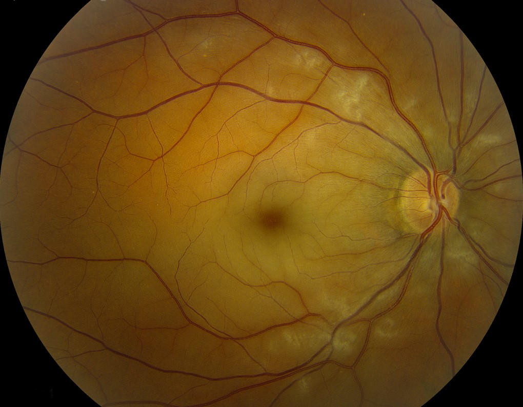
The examiner then sees a tongue of normal-colored retina within the pale edema.
However, the vessel must supply the fovea to preserve good acuity.
Arteritic Disease and Poor CRAO Recovery
Giant cell arteritis can also occlude the posterior ciliary arteries, which adds choroidal and optic nerve ischemia.
As a result, vision rarely improves in the affected eye.
Therefore, ask about headache, jaw claudication and scalp tenderness, then check ESR and CRP urgently.
Corticosteroids mainly aim to protect the fellow eye.
Signs That Predict the Outcome
Initial acuity gives the simplest prognostic signal.
Light perception or worse should raise concern for ophthalmic artery involvement, especially without a cherry red spot.
In addition, a dense relative afferent pupillary defect suggests extensive inner retinal dysfunction.
CRAO Recovery Timeline
Retinal whitening and the cherry red spot dominate the first days.
The whitening then fades over several weeks, and the disc gradually becomes pale.
Any visual improvement tends to appear early, and function usually stabilizes within months.
Therefore, a cleaner-looking fundus does not signal returning vision.
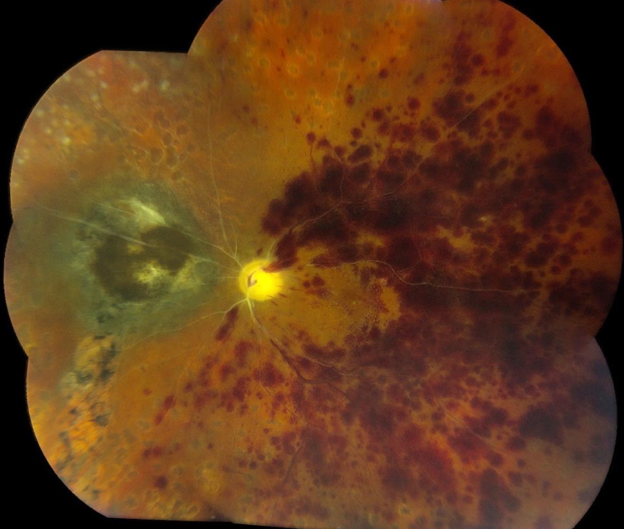
OCT Changes During Follow-Up
Acute OCT shows thickened, hyperreflective inner retinal layers.
Later scans show inner retinal thinning and loss of normal layer definition.
As a result, preserved photoreceptors on OCT should not be read as visual potential.
Can Acute Treatment Improve CRAO Recovery?
Ocular massage, paracentesis and pressure lowering have no proven benefit.
The same meta-analysis found recovery in 50% of patients given intravenous fibrinolysis within 4.5 hours.
However, these data were observational and subject to selection bias.
In the randomized EAGLE trial, intra-arterial fibrinolysis did not outperform conservative treatment and caused more adverse events.
Neovascularization After CRAO
One series reported ocular neovascularization in about 18% of eyes, often within the first few months.
Therefore, examine the undilated iris carefully and perform gonioscopy when suspicion exists.
Panretinal photocoagulation or anti-VEGF therapy may then be needed.
Stroke Risk After the Eye Event
Acute CRAO is a form of ischemic stroke, and vascular risk appears highest soon after onset.
Therefore, patients need same-day referral to a stroke-capable center.
The workup usually includes brain imaging, carotid imaging, cardiac rhythm assessment and risk factor review.
Follow-Up After CRAO
A common approach includes a visit within the first month, then regular checks through about four months.
Each visit should include acuity, pupils, pressure, iris examination and dilated fundus examination.
Next, confirm that the patient has attended stroke prevention follow-up.
Counseling Patients About CRAO Recovery
Start with honest expectations, because many patients hope that vision will return fully.
Explain that the artery may reopen while the damaged nerve cells stay lost.
Also teach urgent warning symptoms, including new loss in either eye, weakness or speech difficulty.
Pitfalls to Avoid
- Do not promise recovery because the fundus has normalized
- Never skip inflammatory markers in an older patient
- Do not stop follow-up after the first visit
- Do not delay stroke evaluation for outpatient imaging


Document what you see
Two smartphone imaging tools built for everyday clinic use — one for the slit lamp, one for the fundus.
From Choroida — the team behind this siteReferences
- Hayreh SS, Zimmerman MB. Central retinal artery occlusion: visual outcome. Am J Ophthalmol. 2005;140:376-391.
- Hayreh SS, Zimmerman MB, Kimura A, Sanon A. Central retinal artery occlusion. Retinal survival time. Exp Eye Res. 2004;78:723-736.
- Schrag M, Youn T, Schindler J, Kirshner H, Greer D. Intravenous fibrinolytic therapy in central retinal artery occlusion: a patient-level meta-analysis. JAMA Neurol. 2015;72:1148-1154.
- Schumacher M, Schmidt D, Jurklies B, et al. Central retinal artery occlusion: local intra-arterial fibrinolysis versus conservative treatment, a multicenter randomized trial. Ophthalmology. 2010;117:1367-1375.
- Rudkin AK, Lee AW, Chen CS. Ocular neovascularization following central retinal artery occlusion: prevalence and timing of onset. Eur J Ophthalmol. 2010;20:1042-1046.
- Mac Grory B, Schrag M, Biousse V, et al. Management of central retinal artery occlusion: a scientific statement from the American Heart Association. Stroke. 2021;52:e282-e294.

Writes and reviews the clinical articles on Educate. Every post is checked against peer-reviewed sources before it goes live.