Silicone oil removal is a planned second operation for many eyes after complex retinal detachment repair.
The decision balances two risks, because oil can damage the eye while removal can expose an unstable retina.
Therefore, surgeons judge each eye individually.
Why Silicone Oil Removal Is Usually Planned
Silicone oil supports the retina by surface tension while chorioretinal adhesion matures.
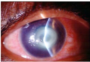
However, prolonged contact raises concern for emulsification, keratopathy, glaucoma and cataract (see silicone oil complications).
Laser or cryotherapy forms the lasting adhesion.
Timing of Silicone Oil Removal
No trial has defined one ideal interval for every eye.
Many surgeons consider removal after about three to six months, although practice varies.
Severe proliferative vitreoretinopathy (PVR) often needs longer observation.
Therefore, the fundus examination should guide the date.
Fundus Explorer Pro
Photograph the retinal findings described here with the phone already in your pocket — 22 D optics and built-in illumination in one handheld unit.
From Choroida — the team behind this siteSigns the Retina Is Ready for Silicone Oil Removal
First, confirm stability through a fully dilated pupil.
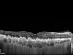
Next, use OCT because oil can obscure subtle macular findings.
Useful signs include:
- An attached retina throughout tamponade
- Mature chorioretinal scars around all breaks
- No progressive star folds or new epiretinal membranes
- No macular traction or subretinal fluid on OCT
Complications That Favor Earlier Removal
Some eyes cannot safely wait.
For example, emulsified droplets may obstruct the trabecular meshwork.
Oil touching the endothelium can also threaten corneal clarity.
In addition, the Silicone Study documented both pressure abnormalities and corneal abnormalities after oil tamponade.
However, removal does not reliably reverse established glaucoma or corneal damage.
Unexplained Vision Loss Under Oil
Some patients report central vision loss while the macula appears attached under oil.
Published reports describe central scotoma developing during tamponade, before removal.
The mechanism remains uncertain, so avoid promising recovery after surgery.
Redetachment Risk After Silicone Oil Removal
Published redetachment rates vary widely between series.
Case mix explains much of this range, because PVR severity and prior surgery differ.
Many recurrences appear within the first months, although later events occur.
Risk Factors for Redetachment
Reported predictors differ among studies, so these factors guide judgment only.
- Severe or recurrent PVR
- Multiple previous repairs
- Large retinectomy or residual traction
- Inferior breaks with incomplete laser coverage
- Open globe trauma and complex tractional disease
Inferior Disease and Silicone Oil Removal
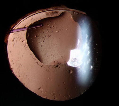
Conventional oil floats, so the inferior retina receives weaker support.
As a result, inferior PVR may progress quietly beneath the bubble.
Therefore, inspect the inferior periphery closely before scheduling removal.
Some eyes need another tamponade (see heavy silicone oil).
When Oil May Stay Long Term
Long-term retention suits some eyes after a frank discussion of risks.
For example, low pressure suggests ciliary body damage, and removal could worsen hypotony.
Repeated redetachment after earlier removal may also favor continued tamponade.
Before Silicone Oil Removal Surgery
First, measure intraocular pressure and compare it with the fellow eye.
Then assess the cornea, gonioscopy findings, lens status and any peripheral iridectomy.
Also, review the original operative note for retinectomy size and laser coverage.
Surgical Technique for Silicone Oil Removal
Most surgeons use small-gauge vitrectomy with active extraction of the oil.
Air-fluid exchange then clears residual droplets from the cavity.
Next, the surgeon inspects the retina, peels residual membranes and adds laser where needed.
Evidence for routine prophylactic laser remains mixed.
Cataract and Silicone Oil Removal
Cataract commonly develops in phakic eyes during tamponade.
Therefore, many surgeons combine lens surgery with removal or schedule it soon afterward (see cataract surgery timing).
Oil alters ultrasound velocity, so optical biometry helps.
Retained oil can also adhere to silicone intraocular lenses, which argues against that material in these eyes.
Vision After Silicone Oil Removal
Refraction changes after removal, particularly in phakic eyes.
Therefore, delay final glasses until the eye stabilizes.
Many patients improve, yet macular damage, PVR and glaucoma may limit the result.
Follow-Up After Silicone Oil Removal
Early visits should check pressure, wound integrity, inflammation and retinal attachment.
Hypotony and pressure spikes can both occur.
Subsequent examinations should include the peripheral retina, because recurrent PVR may start inferiorly.
In addition, OCT helps detect epiretinal membrane and macular edema.
How to Counsel the Patient
Begin by explaining that removal aims to reduce oil-related harm and may improve vision.
Next, state plainly that the retina can detach again.
Patients should also know that vision may stay unchanged or worsen.
Finally, teach patients to report new floaters, flashes or field shadows promptly.
Pitfalls to Avoid
- Removing oil on a fixed date without examining the periphery
- Missing inferior PVR hidden beneath the oil
- Removing oil from a hypotonous eye without discussing phthisis
- Stopping follow-up soon after removal


Document what you see
Two smartphone imaging tools built for everyday clinic use — one for the slit lamp, one for the fundus.
From Choroida — the team behind this siteReferences
- The Silicone Study Group. Vitrectomy with silicone oil or sulfur hexafluoride gas in eyes with severe proliferative vitreoretinopathy: results of a randomized clinical trial. Silicone Study Report 1. Arch Ophthalmol. 1992;110:770-779.
- Barr CC, Lai MY, Lean JS, et al. Postoperative intraocular pressure abnormalities in the Silicone Study. Silicone Study Report 4. Ophthalmology. 1993;100:1629-1635.
- Abrams GW, Azen SP, Barr CC, et al. The incidence of corneal abnormalities in the Silicone Study. Silicone Study Report 7. Arch Ophthalmol. 1995;113:764-769.
- Casswell AG, Gregor ZJ. Silicone oil removal. II. Operative and postoperative complications. Br J Ophthalmol. 1987;71:898-902.
- Jonas JB, Knorr HL, Rank RM, Budde WM. Retinal redetachment after removal of intraocular silicone oil tamponade. Br J Ophthalmol. 2001;85:1203-1207.
- Herbert EN, Habib M, Steel D, Williamson TH. Central scotoma associated with intraocular silicone oil tamponade develops before oil removal. Graefes Arch Clin Exp Ophthalmol. 2006;244:248-252.

Writes and reviews the clinical articles on Educate. Every post is checked against peer-reviewed sources before it goes live.