Elschnig spots are focal choroidal infarcts that appear in severe or accelerated hypertension.
They mark ischemia of the choriocapillaris and the overlying retinal pigment epithelium (RPE). Because they follow a sharp rise in blood pressure, they also carry systemic weight.
What Elschnig Spots Are
Each spot represents a lobule of choriocapillaris that has lost its blood supply, and the RPE above that lobule then becomes ischemic and necrotic. As a result, the lesion lies deep to the retina, at the level of the RPE.
Why the Choroid Fails
Choroidal vessels lack the autoregulation that protects retinal arterioles, so sympathetic tone largely controls choroidal flow and a pressure surge triggers intense vasoconstriction. Fibrinoid necrosis and thrombosis of choroidal arterioles may then occlude the choriocapillaris. Hayreh and colleagues described this sequence in experimental malignant hypertension in monkeys.
Fundus Explorer Pro
Photograph the retinal findings described here with the phone already in your pocket — 22 D optics and built-in illumination in one handheld unit.
From Choroida — the team behind this siteWho Develops Elschnig Spots
Choroidopathy tends to appear when blood pressure rises abruptly, often in younger patients. Sclerotic vessels in long-standing hypertension may partly shield the choroid.
- Malignant hypertension
- Preeclampsia, eclampsia and HELLP syndrome
- Acute renal disease, including scleroderma renal crisis
- Pheochromocytoma
- Thrombotic thrombocytopenic purpura and disseminated intravascular coagulation
Acute Elschnig Spots on Examination
Acute lesions look like small, round, yellow or gray-white patches deep to the retina, and they often cluster in the posterior pole and midperiphery. Overlying retinal vessels cross them without interruption, which confirms their deep location, and numerous acute spots may accompany serous retinal detachment.
Healed Elschnig Spots
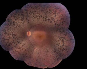
Over several weeks, the acute lesion evolves into a pigmented scar, and the classic healed spot shows a dark pigment clump surrounded by a pale, depigmented halo. Therefore, healed spots record a past hypertensive crisis even when current pressure is normal.
Elschnig Spots on Fluorescein Angiography
In the early phase, acute spots appear hypofluorescent because the affected choriocapillaris does not fill. Later, the lesions stain and may leak as the damaged RPE loses its barrier function. Healed spots behave differently, and their pigmented centers block fluorescence while the atrophic rim shows a window defect.
OCT and ICG Findings
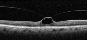
On OCT, acute lesions show hyperreflective changes at the RPE and outer retina, sometimes with subretinal fluid. Healed spots then show RPE irregularity and outer retinal thinning, and indocyanine green angiography can also reveal choroidal hypoperfusion beyond the visible spots.
Siegrist Streaks and Elschnig Spots
Siegrist streaks are linear chains of pigment along sclerosed choroidal arteries. They also signal severe hypertensive choroidopathy, and both signs may coexist in one fundus.

Hypertensive Serous Detachment
Choroidal ischemia damages the RPE pump and the outer blood retinal barrier. As a result, bullous subretinal fluid can mimic central serous chorioretinopathy. However, this fluid typically resolves once blood pressure falls.
What Elschnig Spots Imply About Blood Pressure
Acute spots point to a hypertensive emergency until proven otherwise, and many such eyes also show flame hemorrhages, cotton wool spots, macular star or disc edema. Therefore, the patient needs same-day blood pressure measurement and urgent medical assessment. Healed spots, by contrast, call for review of current control and end-organ status.
Differential Diagnosis in Elschnig Spots
- APMPPE, with placoid lesions and early hypofluorescence
- Serpiginous choroiditis and multifocal choroiditis scars
- Healed toxoplasmosis or histoplasmosis scars
- Pigmented RPE scars after trauma or laser
Blood pressure and pregnancy status usually settle the question.
Workup for Patients With Elschnig Spots
First, measure blood pressure before the patient leaves the clinic, then refer urgently to the emergency department when readings are severely raised. Physicians typically check renal function, urinalysis, blood count and, in pregnancy, liver enzymes and platelets. Young patients also warrant screening for secondary causes of hypertension.
Treatment and Its Limits
No ocular treatment reverses a choroidal infarct. Instead, physicians lower blood pressure in a controlled way, because an abrupt fall risks optic nerve ischemia. Delivery usually resolves the choroidopathy of preeclampsia, and serous detachment then settles while established RPE scars remain.
Visual Prognosis in Elschnig Spots
Vision often recovers well when spots spare the fovea, but macular RPE damage can leave permanent metamorphopsia or reduced acuity. Explain that the scars are stable and reflect systemic risk, so treatment adherence matters. Finally, review the fundus with photographs and OCT a few weeks after pressure control.
Pitfalls to Avoid
- Do not label healed spots as old chorioretinitis without asking about hypertension or pregnancy
- Never send a patient with acute spots home without a blood pressure reading
- Avoid diagnosing central serous chorioretinopathy before checking blood pressure
- Remember that a normal current reading does not exclude a past crisis


Document what you see
Two smartphone imaging tools built for everyday clinic use — one for the slit lamp, one for the fundus.
From Choroida — the team behind this siteReferences
- Hayreh SS, Servais GE, Virdi PS. Fundus lesions in malignant hypertension. VI. Hypertensive choroidopathy. Ophthalmology. 1986;93:1383-1400.
- Tso MO, Jampol LM. Pathophysiology of hypertensive retinopathy. Ophthalmology. 1982;89:1132-1145.
- Wong TY, Mitchell P. Hypertensive retinopathy. N Engl J Med. 2004;351:2310-2317.
- Bowling B. Kanski’s Clinical Ophthalmology: A Systematic Approach. 8th ed. Elsevier; 2016.
- American Academy of Ophthalmology. Basic and Clinical Science Course, Section 12: Retina and Vitreous. San Francisco: AAO.
- Whelton PK, Carey RM, Aronow WS, et al. 2017 guideline for the prevention, detection, evaluation, and management of high blood pressure in adults. Hypertension. 2018;71:e13-e115.
Test yourself
A few questions straight from this article.
-
What does each Elschnig spot represent?
Each spot represents a lobule of choriocapillaris without blood supply; the overlying RPE then becomes ischemic and necrotic, placing the lesion deep to the retina. -
Why is the choroid vulnerable to the pressure surges that cause Elschnig spots?
Choroidal vessels lack the autoregulation that protects retinal arterioles, and sympathetic tone largely controls their flow, so a pressure surge triggers intense vasoconstriction. -
In which setting does the choroidopathy producing Elschnig spots tend to appear?
Choroidopathy tends to appear when blood pressure rises abruptly, often in younger patients, while sclerotic vessels in long-standing hypertension may partly shield the choroid. -
Which examination finding confirms the deep location of acute Elschnig spots?
Overlying retinal vessels cross acute spots without interruption, which confirms that these small yellow or gray-white patches lie deep to the retina. -
What is the classic appearance of a healed Elschnig spot?
Over several weeks the acute lesion becomes a pigmented scar with a dark clump and a pale depigmented halo, recording a past crisis even when current pressure is normal. -
Why do acute Elschnig spots appear hypofluorescent in the early phase of fluorescein angiography?
Early hypofluorescence reflects nonfilling of the affected choriocapillaris; later the lesions stain and may leak as damaged RPE loses its barrier function. -
When does vision often recover well in a patient with Elschnig spots?
Vision often recovers well when spots spare the fovea, whereas macular RPE damage can leave permanent metamorphopsia or reduced acuity. -
Which condition can bullous subretinal fluid from hypertensive choroidopathy mimic?
Choroidal ischemia damages the RPE pump and outer barrier, so the fluid can mimic central serous chorioretinopathy, but it typically resolves once blood pressure falls. -
What does a patient found to have acute Elschnig spots need?
Acute spots point to a hypertensive emergency until proven otherwise, so same-day blood pressure measurement and urgent medical assessment are needed. -
Why is blood pressure lowered in a controlled way in a patient with Elschnig spots?
No ocular treatment reverses a choroidal infarct, so physicians lower blood pressure in a controlled way because an abrupt fall risks optic nerve ischemia.

Writes and reviews the clinical articles on Educate. Every post is checked against peer-reviewed sources before it goes live.