Fleischer ring is a partial or complete ring of iron in the basal corneal epithelium that surrounds the base of the cone in keratoconus.
It appears yellow-brown under white light and dark under cobalt blue light.
Therefore, it helps you map the cone and confirm the diagnosis at the slit lamp.
What the Fleischer Ring Is
Iron deposits form in the basal epithelial cells where the cornea steepens abruptly.
The ring marks the junction between the cone and the flatter surrounding cornea.
Keratoconus is the classic setting, but the ring is not required for the diagnosis, since many patients never develop one.
Because the iron sits in the epithelium, it does not cause symptoms.
How to See the Fleischer Ring
Use the slit lamp with a broad beam and the cobalt blue filter, because contrast is best against a blue background.
The ring may be incomplete, so scan the inferior and temporal cornea first, where the cone often sits.
If the beam is too bright, a faint ring can vanish, so reduce the illumination.
Also, topography maps usually show the steepest area inside the ring.
All-fit Slit-Lamp Adapter
Record and share exactly what you see at the slit lamp. One adapter fits any slit lamp or surgical microscope — and any smartphone.
From Choroida — the team behind this siteFleischer Ring Versus Other Iron Lines
Other iron lines have different positions and causes.
The Hudson-Stahli line is a horizontal line in the lower cornea of older people.
Stocker’s line runs along the head of a pterygium, and Ferry’s line sits in front of a filtering bleb.
Because the Fleischer ring surrounds an ectatic cone, the location and the keratometry make the diagnosis clear (see corneal iron lines).
Fleischer Ring in Practice
The ring does not need treatment.
Instead, it prompts a full ectasia work-up with topography or tomography (see corneal topography).
Progression is judged by keratometry and tomography, not by the ring, so follow those numbers (see keratoconus progression).
Slit-Lamp Tips for the Fleischer Ring
Narrow the beam and widen it again, because a ring seen edge-on can disappear.
Also, add a yellow barrier filter if you have one, since it increases contrast.
Move the beam from the limbus toward the center, and compare both eyes.
Ring Position and Cone Location
The ring marks the base of the cone, so its center usually matches the steepest area on topography.
A nipple cone has a small, central ring, while an oval cone has a larger ring displaced inferiorly.
Because the ring follows the cone, it also shows the extent of the ectasia on a photograph.
Other Signs That Travel With the Ring
Look for other signs of keratoconus in the same eye.
Vogt’s striae, apical thinning, prominent corneal nerves and apical scarring often appear together.
Also, apical scarring at the level of Bowman layer means that the disease is more advanced.
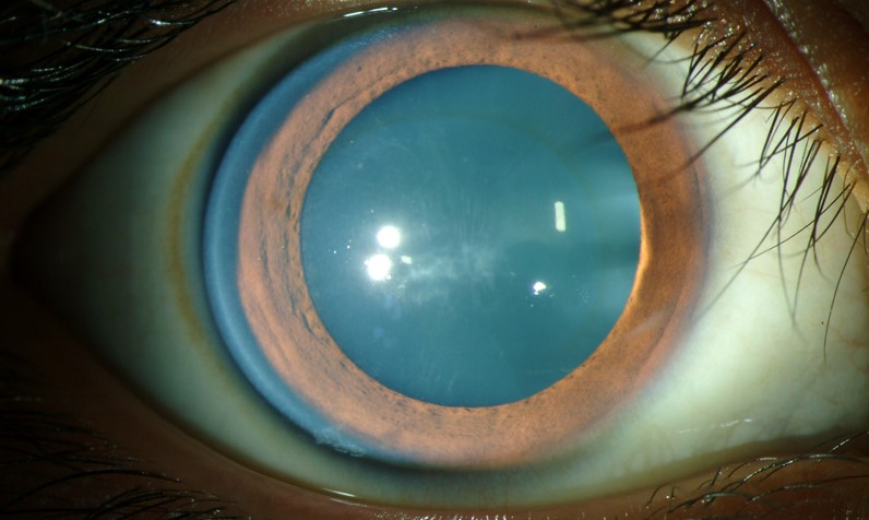
When the Ring Is Absent
A missing ring does not exclude keratoconus.
Early or forme fruste disease may show only subtle topographic changes.
Therefore, keep a high index of suspicion when you see oblique astigmatism or a young patient with a changing refraction.
Counselling and Follow-Up
Explain that the ring is harmless and that it reflects the shape of the cornea.
Also, review the habit of eye rubbing, and treat allergy aggressively, since itch promotes rubbing (see vernal keratoconjunctivitis).
Plan topography every six to twelve months in young patients, and sooner if vision changes.
A Pitfall to Avoid
Do not confuse the ring with the rust ring of a metallic foreign body (see corneal foreign body).
A rust ring sits in the stroma around a fragment and needs removal, but the Fleischer ring needs none.
Also, check the history, because trauma points to a foreign body.
Histology of the Ring
Histologic studies show iron within the basal epithelial cells at the ring.
Therefore, the deposit lies in the epithelium and not in the stroma.
Also, it spares the visual axis, since the ring surrounds the cone.
Theories of Formation
Gass proposed that tear film pooling at a surface irregularity leads to iron deposition in the basal epithelium.
Therefore, the groove around the cone may act as a reservoir.
The exact mechanism is not settled, and the sign carries no prognostic weight.
Associations to Ask About
Keratoconus travels with atopy, Down syndrome, connective tissue disease and floppy eyelid syndrome (see floppy eyelid syndrome).
Also, ask about sleep apnea, since it often accompanies floppy eyelids.
Because these links are common, a short systemic history adds value.
Lens Fitting Around the Ring
The ring marks the edge of the cone, so lens bearing and edge clearance depend on it.
Also, a rigid lens that rests on the ring may cause staining, so review the fit with fluorescein.
For the latest studies, see the PubMed results for Fleischer ring.


Document what you see
Two smartphone imaging tools built for everyday clinic use — one for the slit lamp, one for the fundus.
From Choroida — the team behind this siteReferences
- Rabinowitz YS. Keratoconus. Surv Ophthalmol. 1998;42:297-319.
- Gomes JAP, Tan D, Rapuano CJ, et al. Global consensus on keratoconus and ectatic diseases. Cornea. 2015;34:359-369.
- Gass JDM. The iron lines of the superficial cornea: Hudson-Stahli line, Stocker’s line, and Fleischer’s ring. Arch Ophthalmol. 1964;71:348-358.
- Krachmer JH, Feder RS, Belin MW. Keratoconus and related noninflammatory corneal thinning disorders. Surv Ophthalmol. 1984;28:293-322.

Writes and reviews the clinical articles on Educate. Every post is checked against peer-reviewed sources before it goes live.