
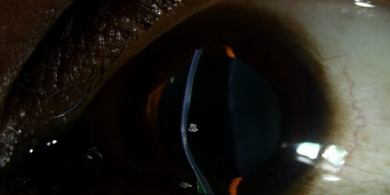
Keratoconus progression is not a subjective clinical impression; it is defined by specific, measurable change in topographic and refractive parameters over a defined period, and having clear criteria matters because corneal crosslinking, the only treatment that halts progression rather than just correcting its optical consequences, is most effective when performed before the cornea has thinned and steepened too far.
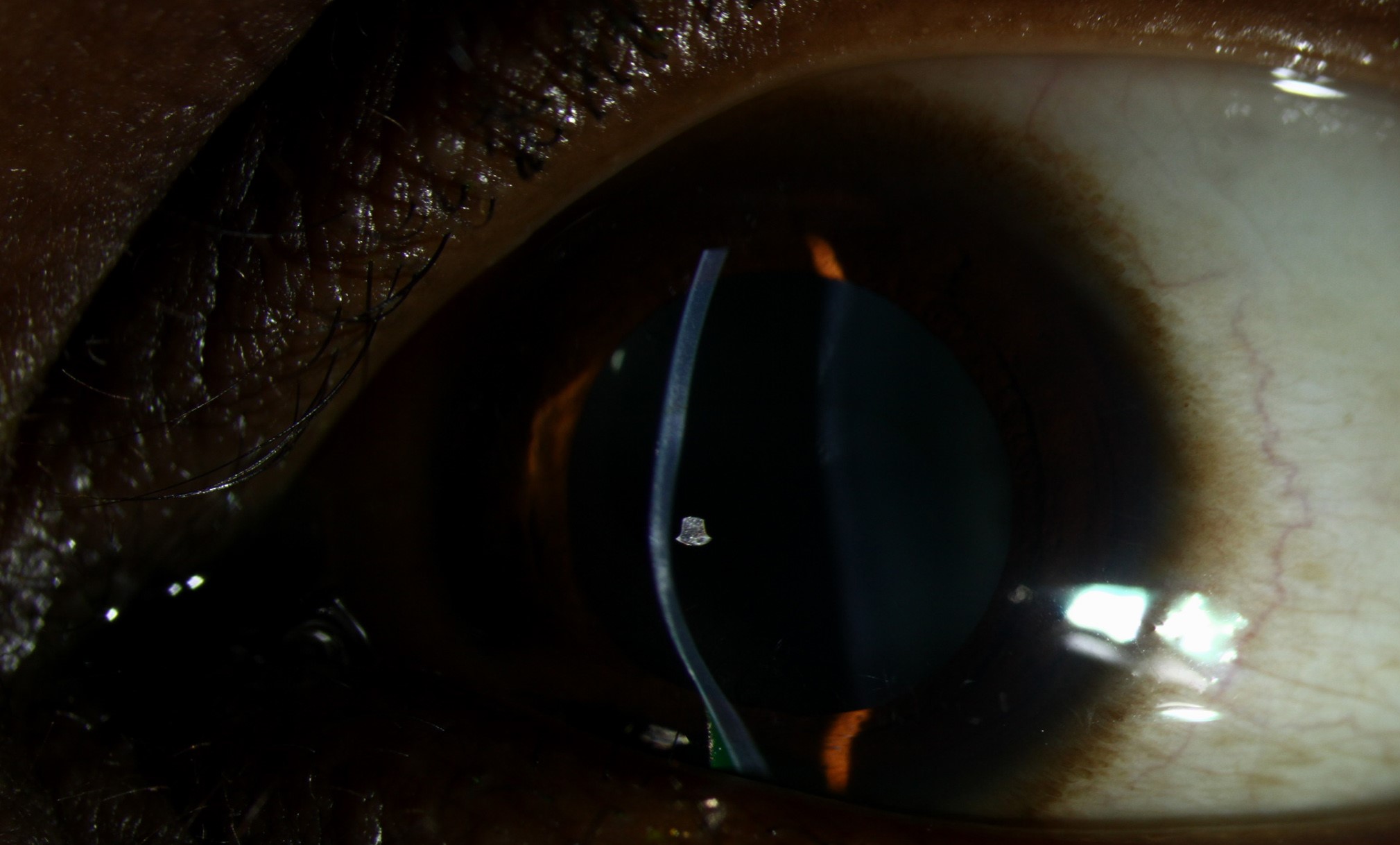
Why Defining Progression Matters
Not every keratoconic cornea progresses, and treating a stable cornea with crosslinking exposes the patient to a procedure’s risks without a clear benefit, while failing to treat a genuinely progressing cornea allows continued, potentially irreversible, structural and optical deterioration.
Consensus criteria for progression give clinicians and patients an objective basis for the crosslinking decision rather than relying on a general sense that the cornea “looks worse.”
Criteria for Progression
The Global Consensus on Keratoconus and Ectatic Diseases and related expert panels have proposed criteria based on change over a defined period, typically 6 to 24 months, using at least two of the following showing consistent change in the direction of worsening ectasia.
- An increase in the steepest keratometry value (Kmax) of 1.0 diopter or more
- A decrease in corneal thickness at the thinnest point of a defined magnitude
- An increase in posterior corneal surface elevation on Scheimpflug or similar tomography
- A myopic shift in manifest refraction, generally a specific increase in spherical equivalent
Different published criteria vary somewhat in the exact thresholds used, but they share the same underlying principle: progression is confirmed by consistent, reproducible change on repeated, quality-controlled measurements, not by a single measurement or subjective impression.
All-fit Slit-Lamp Adapter
Record and share exactly what you see at the slit lamp. One adapter fits any slit lamp or surgical microscope — and any smartphone.
From Choroida — the team behind this siteWhy Repeated, Quality Imaging Matters
Corneal tomography measurements have inherent test-to-test variability, and a single change in Kmax or thickness can reflect measurement noise rather than true progression.
Serial imaging, ideally with the same device and under consistent conditions, and confirmation of change across more than one parameter, reduces the risk of both overtreating a stable cornea based on measurement noise and undertreating a genuinely progressing one because a single ambiguous measurement was dismissed.
Populations at Higher Risk of Progression
- Younger patients, particularly teenagers and young adults, in whom keratoconus tends to progress faster than in older patients, sometimes justifying a lower threshold for treatment or closer monitoring
- Patients with a history of eye rubbing, which is increasingly recognized as a modifiable risk factor for both disease onset and progression
- Patients with atopic disease, since associated eye rubbing and itching contribute to mechanical stress on the cornea
- Down syndrome and certain other conditions associated with keratoconus and eye rubbing behavior
Given the faster typical progression rate in younger patients, some clinicians favor a lower threshold for crosslinking, or even prophylactic treatment of a clearly diagnosed but not yet formally “progressed” cornea, in adolescents, given the high likelihood that progression will eventually be documented anyway (see keratoconus).
Crosslinking Once Progression Is Confirmed
Epithelium-off crosslinking, using riboflavin drops and ultraviolet-A light to increase collagen crosslinking within the corneal stroma, is the standard, most extensively studied technique and has demonstrated durable halting of progression in the large majority of treated eyes in long-term follow-up.
Epithelium-on (transepithelial) techniques aim to reduce the discomfort and epithelial healing time associated with epithelium removal, but evidence for their long-term effectiveness compared with the standard epithelium-off protocol remains less robust, and epithelium-off crosslinking is generally still preferred when maximal treatment effect is the priority.
Monitoring After Crosslinking
Continued topographic and refractive monitoring after crosslinking confirms that progression has stopped, and a small proportion of eyes show continued progression despite treatment and may be considered for repeat crosslinking.
Prognosis
Crosslinking performed before significant corneal thinning and steepening have occurred offers the best chance of preserving useful vision without needing corneal transplantation later, which is why prompt recognition of progression, using objective criteria rather than waiting for obvious clinical deterioration, is central to modern keratoconus management.


Document what you see
Two smartphone imaging tools built for everyday clinic use — one for the slit lamp, one for the fundus.
From Choroida — the team behind this siteReferences
- Gomes JA, Tan D, Rapuano CJ, et al. Global consensus on keratoconus and ectatic diseases. Cornea. 2015;34:359-369.
- Wittig-Silva C, Chan E, Islam FM, Wu T, Whiting M, Snibson GR. A randomized, controlled trial of corneal collagen cross-linking in progressive keratoconus: three-year results. Ophthalmology. 2014;121:812-821.
- Wollensak G, Spoerl E, Seiler T. Riboflavin/ultraviolet-a-induced collagen crosslinking for the treatment of keratoconus. Am J Ophthalmol. 2003;135:620-627.
- Duncan JK, Belin MW, Borgstrom M. Assessing progression of keratoconus: novel tomographic determinants. Eye Vis (Lond). 2016;3:6.
