
A retinal pigment epithelium window defect is a specific fluorescein angiography finding, not a diagnosis in itself, and understanding exactly what it represents, missing or attenuated pigment simply allowing background choroidal fluorescence to shine through more visibly, is essential for correctly interpreting angiograms and avoiding confusion with the other, more clinically active causes of hyperfluorescence that this pattern must be distinguished from.
What a Window Defect Actually Represents
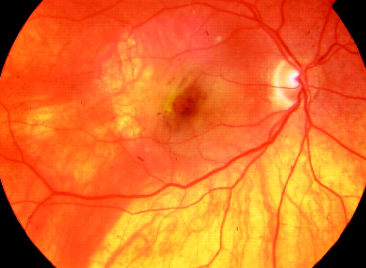
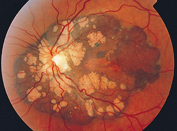
The retinal pigment epithelium normally contains melanin pigment that partially blocks and masks the fluorescence of dye within the underlying choroidal circulation during fluorescein angiography, and when this pigment epithelium becomes thinned, atrophic, or entirely absent, this normal masking effect is lost, allowing the underlying choroidal fluorescence to become more visible, a phenomenon termed a window defect.
The Key Distinguishing Angiographic Behavior
Window defects have a characteristic and diagnostically essential temporal pattern that separates them from other causes of hyperfluorescence: they appear early in the angiogram, corresponding to the timing of the underlying choroidal fluorescence itself, remain sharply demarcated with well-defined borders corresponding exactly to the area of pigment epithelial loss, and, critically, do not increase in size or intensity, and do not leak or pool, as the angiogram progresses through its later frames.
This lack of leakage or progressive change over the course of the angiogram is what distinguishes a window defect from other causes of hyperfluorescence, including active leakage from choroidal neovascularization or staining of an anatomic structure, both of which characteristically increase in size, intensity, or extent in later angiogram frames.
Fundus Explorer Pro
Photograph the retinal findings described here with the phone already in your pocket — 22 D optics and built-in illumination in one handheld unit.
From Choroida — the team behind this siteCauses of Window Defects
- Geographic atrophy in advanced age-related macular degeneration, one of the most common causes, where areas of retinal pigment epithelial atrophy produce well-demarcated window defects corresponding precisely to the atrophic areas
- Pattern dystrophies and other inherited retinal pigment epithelial conditions producing focal areas of pigment epithelial thinning
- Resolved chorioretinitis or other inflammatory or infectious processes leaving behind an area of pigment epithelial atrophy or scarring
- Any other cause of focal or diffuse retinal pigment epithelial atrophy or attenuation, including some inherited retinal degenerations
Distinguishing From Other Angiographic Patterns
- Leakage from choroidal neovascularization shows progressively increasing hyperfluorescence with indistinct, blurring borders in later frames, in clear contrast to the static, sharply demarcated appearance of a window defect
- Pooling, as seen with subretinal fluid in central serous chorioretinopathy or other conditions, shows fluorescence accumulating within a defined anatomic space, typically increasing in intensity and sometimes changing shape over the course of the angiogram
- Staining, as seen with drusen or scar tissue, shows a gradual increase in fluorescence intensity within the fixed boundaries of the anatomic structure, without the leakage pattern of active neovascularization but also without the fully static behavior of a pure window defect
Clinical Use
Recognizing a window defect for what it is, simple absence of normal pigment masking rather than any form of active leakage or abnormal fluid accumulation, prevents misinterpreting a benign or stable atrophic process as active neovascular or inflammatory disease requiring urgent treatment.
Correctly identifying window defects is particularly important when evaluating a patient with age-related macular degeneration for choroidal neovascularization, since correctly distinguishing pure geographic atrophy (producing window defects) from neovascular disease (producing progressive leakage) directly determines whether anti-VEGF treatment is indicated.
Correlation With Other Imaging
Optical coherence tomography, showing direct structural evidence of retinal pigment epithelial thinning or loss corresponding to the angiographic window defect, provides complementary structural confirmation and is now often used alongside or even in place of fluorescein angiography for characterizing geographic atrophy in routine clinical practice.
Clinical Significance
Understanding the window defect pattern is fundamental angiographic literacy, since correctly distinguishing this benign, non-leaking pattern from the various forms of pathologic leakage and staining directly shapes treatment decisions across a wide range of retinal conditions, from age-related macular degeneration to inflammatory chorioretinal disease.


Document what you see
Two smartphone imaging tools built for everyday clinic use — one for the slit lamp, one for the fundus.
From Choroida — the team behind this siteReferences
- Gass JD. Stereoscopic Atlas of Macular Diseases: Diagnosis and Treatment. 4th ed. St. Louis: Mosby; 1997.
- Yannuzzi LA. The Retinal Atlas. 2nd ed. Edinburgh: Elsevier; 2017.
- Bird AC, Bressler NM, Bressler SB, et al. An international classification and grading system for age-related maculopathy and age-related macular degeneration. Surv Ophthalmol. 1995;39:367-374.
- Holz FG, Bindewald-Wittich A, Fleckenstein M, et al. Progression of geographic atrophy and impact of fundus autofluorescence patterns in age-related macular degeneration. Am J Ophthalmol. 2007;143:463-472.
