
Corneal topography maps the curvature of the corneal surface, producing a colour-coded representation that reveals irregularities invisible to keratometry or a slit-lamp examination alone.
Its clinical value is concentrated in a handful of applications, screening for subclinical keratoconus before refractive surgery, monitoring ectatic disease, and planning contact lens fits and toric intraocular lenses, but within those applications it is close to indispensable.
How Corneal Topography Works
Placido-disc based systems, the original and still widely used technology, project a series of concentric illuminated rings onto the cornea and analyse the reflected pattern. An irregular cornea distorts the spacing of these rings, and software converts that distortion into a curvature map.
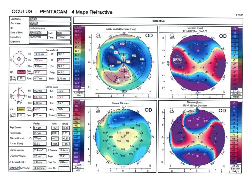
Scanning-slit and Scheimpflug-based systems, in contrast, capture both the anterior and posterior corneal surfaces directly, generating elevation-based maps rather than relying purely on reflected ring analysis. This posterior surface data is something Placido-based systems cannot provide, which is a meaningful advantage of Scheimpflug technology, particularly for detecting subtle ectatic change.
Output is typically displayed as an axial or sagittal curvature map, colour-coded from cool colours for flatter areas to warm colours for steeper ones, alongside numerical indices summarising overall corneal shape and regularity.
Clinical Applications
Refractive Surgery Screening
Identifying subclinical or forme fruste keratoconus before LASIK or PRK is arguably the single most important use of corneal topography in everyday practice, since operating on an unrecognised ectatic cornea risks inducing post-refractive ectasia, one of the most serious complications in refractive surgery.
Keratoconus Diagnosis and Monitoring
Topography reveals the characteristic patterns of keratoconus, typically inferior or central steepening, well before the disease is visible on slit-lamp examination, and serial topography over time is used to document progression, which is itself the trigger for considering corneal cross-linking.
Contact Lens Fitting
Detailed corneal shape data supports fitting of specialty contact lenses, including scleral and rigid gas-permeable lenses, particularly in irregular corneas where a standard lens fit would be unpredictable.
Toric Intraocular Lens Planning
Accurate measurement of corneal astigmatism, including its axis, is essential for selecting and correctly orientating a toric intraocular lens at the time of cataract surgery, and topography contributes to this measurement alongside keratometry.
All-fit Slit-Lamp Adapter
Record and share exactly what you see at the slit lamp. One adapter fits any slit lamp or surgical microscope — and any smartphone.
From Choroida — the team behind this siteInterpreting Common Patterns
- Regular astigmatism appears as a symmetric bowtie pattern, generally not a cause for concern on its own
- Asymmetric bowtie or inferior steepening patterns raise suspicion for keratoconus or pellucid marginal degeneration and warrant closer clinical correlation
- Irregular, non-orthogonal patterns can indicate corneal scarring, prior surgery, or significant dry eye affecting the tear film over which the measurement is taken
- A poor-quality or unreliable scan, often from tear film irregularity or poor fixation, should prompt a repeat measurement rather than acting on unreliable data
Tear film quality genuinely matters here: dry eye or an unstable tear film can produce topographic irregularity that mimics true corneal pathology, which is why the ocular surface should be optimised before a topography scan whenever possible, particularly ahead of refractive surgery planning.
Limitations
Placido-based systems measure only the anterior corneal surface, missing posterior surface changes that can be an early sign of ectasia, which is a meaningful reason Scheimpflug-based tomography has become preferred for refractive surgery screening in many practices.
Topography alone does not diagnose keratoconus; it is interpreted alongside clinical examination, pachymetry, and, where available, posterior elevation data, since topographic irregularity has several possible causes beyond ectatic disease.
Poor image quality from blinking, dry eye, or poor patient fixation is a common practical limitation, and results should always be checked for scan quality before being used for a clinical decision.
Prognosis and Clinical Impact
As a screening tool, corneal topography has measurably reduced the incidence of post-refractive ectasia by identifying at-risk corneas before surgery, which is probably its single greatest contribution to modern ophthalmic practice.
In keratoconus management, serial topography is what defines progression and therefore determines the timing of cross-linking, directly shaping the visual outcome of that treatment by supporting earlier intervention.
Its role continues to expand alongside newer Scheimpflug and OCT-based tomography systems, which add posterior corneal and pachymetric data to the surface curvature information that Placido-based topography alone provides.


Document what you see
Two smartphone imaging tools built for everyday clinic use — one for the slit lamp, one for the fundus.
From Choroida — the team behind this siteReferences
- Rabinowitz YS. Videokeratographic indices to differentiate normal and keratoconic eyes. Journal of Refractive Surgery. 1995.
- Belin MW, Khachikian SS. An introduction to understanding elevation-based topography: how elevation data is displayed – a review. Clinical and Experimental Ophthalmology. 2009.
- Randleman JB, Trattler WB, Stulting RD. Validation of the Ectasia Risk Score System for preoperative laser in situ keratomileusis screening. American Journal of Ophthalmology. 2008.
- Corneal Topography. EyeWiki, American Academy of Ophthalmology.
- Corneal Topography. StatPearls, NCBI Bookshelf.
Test yourself
A few questions straight from this article.
-
What does corneal topography reveal that keratometry or slit-lamp examination alone cannot?
Topography maps surface curvature as a colour-coded display, exposing irregularities that keratometry or a slit-lamp view would miss. -
How does a Placido-disc topographer derive a curvature map?
Concentric illuminated rings are projected onto the cornea, and software converts the distortion in their reflected spacing into a curvature map. -
What key data do Scheimpflug-based systems provide that Placido-disc topography cannot?
Scheimpflug and scanning-slit systems capture both corneal surfaces, and that posterior elevation data is a meaningful advantage for detecting subtle ectatic change. -
On a standard corneal curvature map, what do warm colours represent?
Axial or sagittal maps are colour-coded with cool colours for flatter regions and warm colours for steeper ones. -
Which complication does preoperative corneal topography before LASIK or PRK chiefly aim to prevent?
Detecting subclinical or forme fruste keratoconus before surgery is topography's most important everyday role, because operating on an unrecognised ectatic cornea risks post-refractive ectasia. -
Which topographic pattern typically raises suspicion of keratoconus?
Inferior or central steepening and asymmetric bowtie patterns suggest keratoconus or pellucid marginal degeneration and warrant close clinical correlation. -
A symmetric bowtie pattern on corneal topography most often represents which finding?
A symmetric bowtie reflects regular astigmatism and is generally not a cause for concern on its own. -
Why should the ocular surface be optimised before a corneal topography scan?
Dry eye or an unstable tear film produces topographic irregularity that can imitate genuine corneal disease, which matters most before refractive surgery planning. -
In keratoconus, what finding on serial corneal topography triggers consideration of cross-linking?
Serial topography is what defines progression, and progression is the trigger for considering corneal cross-linking, so timing of treatment depends on it. -
Which statement reflects a genuine limitation of corneal topography?
Topographic irregularity has several possible causes, so maps must be read alongside clinical examination, pachymetry and, where available, posterior elevation data.

Writes and reviews the clinical articles on Educate. Every post is checked against peer-reviewed sources before it goes live.


