Giant cell arteritis is a vasculitis of large and medium arteries that can cause sudden, permanent blindness.
Vision loss may be the first sign and may affect the second eye within days.
Therefore, suspect it in every older patient with new visual symptoms.
Who Gets Giant Cell Arteritis
It affects people over fifty and is more common in women.
Polymyalgia rheumatica accompanies a share of cases.
Also, patients may have headache, jaw claudication and scalp tenderness.
Because symptoms vary, ask about each of them.
Ocular Presentations
Arteritic anterior ischemic optic neuropathy is the most common cause of vision loss (see arteritic anterior ischemic optic neuropathy).
Central retinal artery occlusion and cilioretinal artery occlusion also occur (see central retinal artery occlusion).
Also, amaurosis fugax and diplopia may precede vision loss (see amaurosis fugax).
Therefore, transient symptoms are warnings.
Fundus Explorer Pro
Photograph the retinal findings described here with the phone already in your pocket — 22 D optics and built-in illumination in one handheld unit.
From Choroida — the team behind this siteOccult Giant Cell Arteritis
Hayreh and colleagues described patients without systemic symptoms who had ocular disease.
These patients had occult disease and were at risk of delay.
Also, a normal history does not exclude the diagnosis.
Because the risk is high, test every older patient with arteritic features.
Findings on Examination in Giant Cell Arteritis
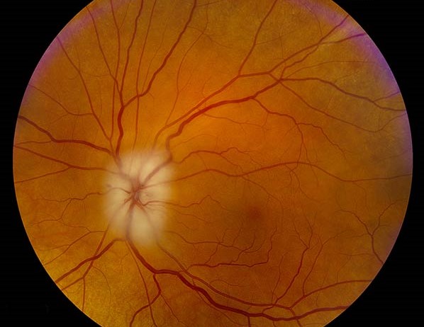
The disc often shows pallor with chalky white edema.
The pupil shows a relative afferent defect.
Also, vision is often poor, with counting fingers or worse (a relative afferent pupillary defect is typical).
Therefore, the picture differs from the nonarteritic form.
Distinguishing From Nonarteritic Disease
Nonarteritic ischemic optic neuropathy shows a crowded disc and milder loss (see nonarteritic ischemic optic neuropathy).
Giant cell arteritis shows pale swelling and a high inflammatory marker level.
Also, fluorescein shows delayed choroidal filling in arteritic disease.
Because management differs, make the distinction early.
Blood Tests
Order the erythrocyte sedimentation rate and the C-reactive protein at once.
A raised platelet count supports the diagnosis.
Also, no single test excludes the disease.
Therefore, treat the patient based on the clinical picture.
Starting Steroids Immediately in Giant Cell Arteritis
Start high-dose corticosteroids as soon as you suspect the diagnosis.
In patients with visual symptoms, many centers give intravenous methylprednisolone for three days.
Also, oral prednisone follows at a high dose.
Because vision loss is permanent, do not wait for biopsy.
Temporal Artery Biopsy for Giant Cell Arteritis
Biopsy confirms the diagnosis and should be done within days to a couple of weeks of starting steroids.
A long specimen reduces the chance of missing skip lesions.
Also, a negative biopsy does not exclude the disease in strong clinical cases.
Therefore, interpret the result with the picture.
Ultrasound of the Temporal Arteries
The halo sign is a dark rim around the artery lumen.
The TABUL study compared ultrasound with biopsy.
Also, ultrasound is quick and noninvasive.
Because it is operator dependent, use it with experienced staff.
Steroid-Sparing Therapy in Giant Cell Arteritis
Tocilizumab reduced relapses and steroid exposure in the GiACTA trial.
Methotrexate has smaller data.
Also, long steroid courses cause harm.
Therefore, involve rheumatology early.
Risk to the Second Eye
Untreated patients often lose vision in the second eye within days to weeks.
Hayreh and Zimmerman reported visual loss despite high-dose steroids in some patients.
Also, prompt treatment lowers the risk.
Because the consequences are severe, act without delay.
Counseling
Explain that the disease is serious and that treatment is long.
Also, warn about steroid side effects and monitoring.
Because patients need support, arrange regular follow-up.
Systemic Symptoms to Ask About
Ask about headache, scalp tenderness, jaw pain on chewing and weight loss.
Also, ask about fever, fatigue and shoulder or hip stiffness.
Because patients may not link these symptoms to the eye, ask directly.
Differential Diagnosis
Other causes of vision loss in older adults include nonarteritic ischemic neuropathy, retinal vein occlusion and embolic events.
Also, infectious and inflammatory causes occur.
Therefore, use the inflammatory markers to prioritize.
Steroid Dosing and Taper
Typical oral doses start near one milligram per kilogram of prednisone.
The taper takes many months and follows symptoms and markers.
Also, add bone protection and monitor glucose and blood pressure.
Because relapse occurs, taper slowly.
Follow-Up and Long-Term Care
Check inflammatory markers and symptoms during the taper.
Also, watch for aortic involvement later in the course.
Therefore, coordinate with rheumatology for long-term care.
For the latest studies, see the PubMed results for giant cell arteritis.


Document what you see
Two smartphone imaging tools built for everyday clinic use — one for the slit lamp, one for the fundus.
From Choroida — the team behind this siteReferences
- Hayreh SS, Podhajsky PA, Zimmerman B. Occult giant cell arteritis: ocular manifestations. Am J Ophthalmol. 1998;125:521-526.
- Hunder GG, Bloch DA, Michel BA, et al. The American College of Rheumatology 1990 criteria for the classification of giant cell arteritis. Arthritis Rheum. 1990;33:1122-1128.
- Stone JH, Tuckwell K, Dimonaco S, et al. Trial of tocilizumab in giant-cell arteritis. N Engl J Med. 2017;377:317-328.
- Luqmani R, Lee E, Singh S, et al. The role of ultrasound compared to biopsy of temporal arteries in the diagnosis and treatment of giant cell arteritis (TABUL): a diagnostic accuracy and cost-effectiveness study. Health Technol Assess. 2016;20:1-238.

Writes and reviews the clinical articles on Educate. Every post is checked against peer-reviewed sources before it goes live.