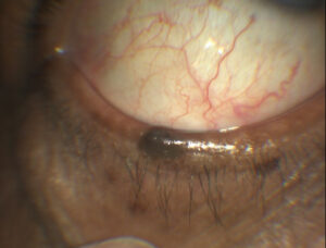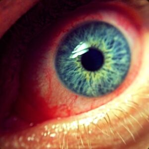
Floppy eyelid syndrome is a condition of abnormally lax, rubbery upper eyelids that evert easily — sometimes spontaneously during sleep — producing chronic mechanical irritation of the ocular surface.
It is strongly, though not universally, associated with obstructive sleep apnea and obesity, and is one of the more commonly overlooked causes of chronic red eye and papillary conjunctivitis in middle-aged and older men, in part because the diagnostic finding (an abnormally lax upper lid) has to be specifically tested for rather than being obvious on routine inspection.
Association With Sleep Apnea
The link between floppy eyelid syndrome and obstructive sleep apnea is well established, and the proposed mechanism involves mechanical trauma from the lid rubbing against the pillow or bedding during sleep, compounded in some patients by tissue changes related to intermittent hypoxia from apneic episodes affecting the eyelid’s elastin content.
Given this association, patients diagnosed with floppy eyelid syndrome — those with a body habitus or symptoms (snoring, daytime fatigue) suggestive of sleep apnea — warrant screening for sleep apnea, because identifying and treating the sleep disorder can be as important to overall health as managing the eye findings themselves.
This makes floppy eyelid syndrome one of a small number of ophthalmic findings that functions as a genuine systemic screening opportunity, since an ophthalmologist recognizing the lid laxity may be the first clinician to prompt a sleep study in a patient who had never previously been evaluated for apnea.
Pathogenesis
Histologically, the tarsal plate in affected eyelids shows a marked reduction in elastin content, which is thought to underlie the abnormal laxity and the tendency of the lid to evert with even minor mechanical stress.
Patients characteristically sleep in a position that allows the upper lid to spontaneously evert against the pillow, exposing the palpebral conjunctiva to prolonged mechanical friction throughout the night without the patient being aware it is happening.
Whether the reduced elastin is a primary connective tissue abnormality or a secondary consequence of repeated mechanical and hypoxic stress over years remains debated, and it is possible that both mechanisms reinforce one another once the process has begun, with early laxity increasing mechanical exposure and mechanical exposure further degrading the tissue.
All-fit Slit-Lamp Adapter
Record and share exactly what you see at the slit lamp. One adapter fits any slit lamp or surgical microscope — and any smartphone.
From Choroida — the team behind this siteClinical Presentation
Patients typically present with chronic, nonspecific ocular irritation, redness, mucous discharge, and a foreign-body sensation that is often worse in the morning upon waking — a pattern that should specifically raise suspicion for floppy eyelid syndrome, because the overnight mechanical exposure is the primary driver of symptoms.
Many patients have been treated for presumed allergic or bacterial conjunctivitis for a considerable time before the diagnosis is actually made, given how nonspecific the symptoms are without a targeted lid exam.
Asking specifically about which side the patient habitually sleeps on can be a useful clue, since floppy eyelid syndrome is often asymmetric or unilateral, corresponding to the side pressed against the pillow most consistently during sleep.
Exam Findings
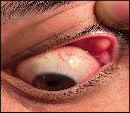
- Easily everted upper eyelid with minimal upward traction — the key diagnostic maneuver, testing lid laxity by gently pulling the upper lid upward and observing how readily it flips
- A rubbery, soft texture to the tarsal plate on palpation
- Papillary conjunctivitis on the everted superior tarsal conjunctiva, from chronic mechanical irritation
- Punctate epithelial staining, sometimes superior in distribution similar to other conditions involving superior corneal exposure or friction
- Ptosis and lash ptosis are common associated findings, related to the same underlying tissue laxity
Differential Diagnosis
- Superior limbic keratoconjunctivitis — superior bulbar and limbal findings from a different mechanical mechanism (redundant bulbar conjunctiva rather than lid laxity), discussed in its own dedicated article on this site
- Chronic allergic or giant papillary conjunctivitis — tarsal papillae from allergen or contact lens exposure rather than eyelid laxity, without the characteristic easy eversion on exam
- Chronic blepharitis — lid margin disease without the specific finding of abnormal tarsal laxity
The single most useful step in reaching the correct diagnosis is remembering to test upper lid laxity in any patient with chronic, otherwise unexplained conjunctival irritation, especially in an overweight, middle-aged or older man with symptoms worse in the morning.
Diagnostic Evaluation
The diagnosis is made clinically at the bedside, by gently grasping and lifting the upper lid margin and observing how easily it everts compared with the expected resistance of a normal, appropriately taut lid.
Slit-lamp exam of the everted tarsal conjunctiva documents the degree of papillary change and any associated punctate staining.
Because the association with sleep apnea is so clinically important, a directed sleep history — snoring, witnessed apneic episodes, excessive daytime sleepiness — should be part of the standard workup once floppy eyelid syndrome is diagnosed, with formal polysomnography referral for patients whose history is suggestive.
Management
Nighttime protective measures — taping the eyelid closed, a shield, or an eye mask — reduce mechanical exposure during sleep and are a reasonable first-line, low-risk intervention, alongside lubrication to address the ocular surface irritation.
Referral for sleep apnea evaluation and treatment (often with continuous positive airway pressure) is appropriate for patients with suggestive symptoms or risk factors, both for the systemic health benefits and because effective sleep apnea treatment may indirectly improve the eyelid findings for some patients.
Surgical lid-tightening (horizontal lid shortening, similar in concept to procedures used for involutional entropion or ectropion) is reserved for patients with persistent, significant symptoms despite conservative measures, and directly addresses the underlying tissue laxity that conservative management cannot correct.


Document what you see
Two smartphone imaging tools built for everyday clinic use — one for the slit lamp, one for the fundus.
From Choroida — the team behind this siteReferences
- Culbertson WW, Ostler HB. The floppy eyelid syndrome. American Journal of Ophthalmology.
- McNab AA. The eye and sleep apnea. Sleep Medicine Reviews.
- Ezra DG, Beaconsfield M, Collin R. Floppy eyelid syndrome: stretching the limits. Survey of Ophthalmology.
- American Academy of Ophthalmology. Basic and Clinical Science Course, Section 7: Orbit, Eyelids, and Lacrimal System.
Test yourself
A few questions straight from this article.
-
Which eyelid abnormality defines floppy eyelid syndrome?
The lids are abnormally lax and evert with minimal force, sometimes spontaneously during sleep, producing chronic mechanical irritation of the ocular surface. -
Which systemic condition is strongly associated with floppy eyelid syndrome?
The association with obstructive sleep apnea and obesity is well established, and recognising the lid laxity may be what first prompts a sleep study. -
What histological change is found in the tarsal plate in floppy eyelid syndrome?
Reduced tarsal elastin is thought to underlie the abnormal laxity and the tendency of the lid to evert under even minor mechanical stress. -
Which bedside manoeuvre establishes the diagnosis of floppy eyelid syndrome?
Gently grasping and lifting the upper lid, and comparing how easily it everts with the resistance of a normal taut lid, is the key diagnostic test and must be actively performed. -
Why are the ocular symptoms of floppy eyelid syndrome typically worst on waking?
The lid everts against the pillow during sleep, exposing the palpebral conjunctiva to hours of friction, so irritation, redness and discharge are worst on waking. -
Once floppy eyelid syndrome is diagnosed, which step belongs in the standard workup?
Asking about snoring, witnessed apneas and daytime sleepiness, and referring suggestive patients for a sleep study, is part of the standard workup given how important the apnea link is. -
Which conjunctival finding results from the chronic friction in floppy eyelid syndrome?
Chronic mechanical irritation of the everted upper lid produces papillary change on the superior tarsal conjunctiva, seen once the lid is everted at the slit lamp. -
Which mechanism separates superior limbic keratoconjunctivitis from floppy eyelid syndrome?
Superior limbic keratoconjunctivitis produces superior bulbar and limbal findings through redundant bulbar conjunctiva, a different mechanical mechanism from lid laxity. -
What is reasonable first-line management for floppy eyelid syndrome?
Taping the lid closed, a shield or an eye mask reduces overnight mechanical exposure and is a low-risk first step, with lubrication for the surface irritation. -
When is surgical lid-tightening indicated in floppy eyelid syndrome?
Horizontal lid shortening directly addresses the tissue laxity that conservative treatment cannot correct, and is reserved for patients still symptomatic after those measures.

Writes and reviews the clinical articles on Educate. Every post is checked against peer-reviewed sources before it goes live.