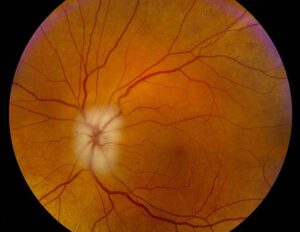
Fat embolism retinopathy is a retinal complication of fat embolism syndrome after trauma or surgery.
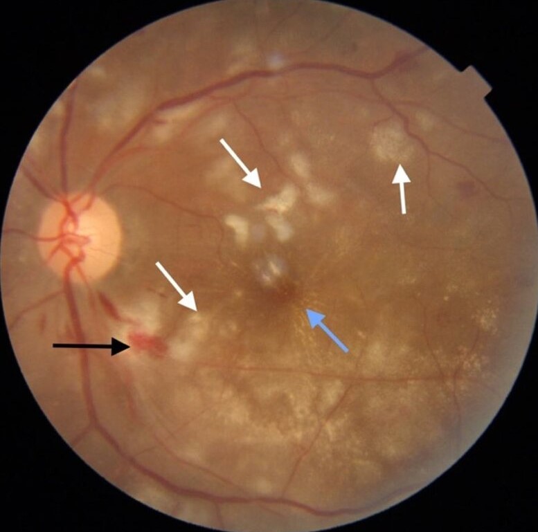
It shows cotton-wool spots and hemorrhages around the disc and macula.
Therefore, it should be considered in patients with fractures who report new visual symptoms.
What Fat Embolism Is
Fat globules enter the circulation after long bone fracture or orthopedic surgery.
They lodge in small vessels of the lung, brain, skin and retina.
Also, they trigger an inflammatory response.
As a result, the syndrome affects many organs.
When Fat Embolism Syndrome Occurs
Symptoms start 12 to 72 hours after injury.
Femoral and tibial fractures are the main causes.
Also, intramedullary nailing and joint replacement carry risk.
Therefore, timing helps to identify the syndrome.
Fundus Explorer Pro
Photograph the retinal findings described here with the phone already in your pocket — 22 D optics and built-in illumination in one handheld unit.
From Choroida — the team behind this siteClassic Features
Gurd described the clinical criteria in 1970.
Major signs include respiratory distress, neurological changes and a petechial rash.
Also, minor signs include fever, tachycardia and retinal changes.
In addition, anemia and low platelets are common.
Retinal Findings in Fat Embolism Retinopathy
The fundus shows cotton-wool spots, intraretinal hemorrhages and sometimes macular edema.
Lesions cluster around the disc and posterior pole.
Also, many patients have no visual symptoms.
As a result, the retinal signs may be found only on examination.
Symptoms of Fat Embolism Retinopathy
Patients may report blurred vision or scotomas.
Sedated patients cannot report symptoms.
Also, central loss occurs when the macula is involved.
Therefore, examine patients at risk who cannot speak.
Mechanism of Fat Embolism Retinopathy
Fat emboli block the precapillary arterioles.
They cause local ischemia and release of free fatty acids.
Also, endothelial injury leads to leakage.
As a result, the picture resembles other embolic retinopathies.
Comparison with Purtscher Retinopathy
Purtscher retinopathy occurs after chest compression or head trauma.
Agrawal and McKibbin reviewed the Purtscher and Purtscher-like entities.
Also, fat embolism is one cause of a Purtscher-like picture.
Therefore, the term Purtscher-like fits many cases.
Other Causes of Purtscher-Like Retinopathy
Pancreatitis, renal failure and lupus can cause the same fundus.
Childbirth and connective tissue disease are also known causes.
Also, complement activation is a shared mechanism.
In addition, a careful history separates the causes.
Differential Diagnosis
Diabetic and hypertensive retinopathy produce similar spots.
Anemia, leukemia and sepsis also cause cotton-wool spots.
Also, talc and other embolic retinopathy occur in drug users.
Therefore, consider the clinical setting.
Investigations for Fat Embolism Retinopathy
Dilated fundus examination is the key test.
OCT shows inner retinal thickening and sometimes outer retinal changes.
Also, fluorescein angiography shows areas of nonperfusion.
As a result, imaging maps the damage.
Systemic Management
Treatment is supportive and focuses on oxygen and fluids.
Early fracture fixation lowers risk.
Also, steroids have been tried without clear proof of benefit.
Therefore, systemic care leads.
Ocular Management of Fat Embolism Retinopathy
No specific treatment exists for the retinal lesions.
Observe most eyes and treat complications.
Also, anti-VEGF drugs help macular edema or neovascularization in rare cases.
In addition, follow with OCT.
Prognosis of Fat Embolism Retinopathy
Most retinal lesions resolve over weeks to months.
Vision recovers in many patients.
Also, macular involvement and nonperfusion lead to lasting loss.
As a result, early examination sets expectations.
Relation to Brain Findings
Brain emboli cause confusion, seizures and coma.
Retinal signs may parallel brain involvement.
Also, MRI shows a starfield pattern of small lesions.
Therefore, the eye can reflect the brain.
Prevention
Stabilize fractures early.
Use careful surgical technique to limit marrow release.
Also, monitor high-risk patients in the first days.
In addition, involve the orthopedic team in care.
Pitfalls to Avoid
- Do not ignore a fundus examination in an intubated patient with a long bone fracture
- Never label cotton-wool spots as diabetic without a history
- Do not expect steroids to reverse the retinal lesions
- Never forget to follow macular lesions with OCT
Systemic Evaluation
Patients need oxygen monitoring, blood counts and brain imaging when symptoms suggest involvement.
A skin and conjunctival check can show petechiae.
Also, check for low platelets.
Therefore, the eye exam fits into a wider evaluation.
Conjunctival and Skin Findings
Petechiae on the conjunctiva, chest and axillae are classic.
They appear a day or two after injury.
Also, they fade within days.
As a result, they provide a quick clue.
Role of Retinal Imaging
OCT shows thickening of the inner retina at cotton-wool spots.
Angiography shows capillary nonperfusion.
Also, the images help to follow recovery.
In addition, widefield imaging shows peripheral lesions.
Follow-Up Plan
Examine the eyes after the acute illness and again in a month.
Repeat OCT if vision is reduced.
Also, refer patients with persistent scotomas.
Therefore, plan a clear schedule.
For the latest studies, see the PubMed results for fat embolism retinopathy.


Document what you see
Two smartphone imaging tools built for everyday clinic use — one for the slit lamp, one for the fundus.
From Choroida — the team behind this siteReferences
- Gurd AR. Fat embolism: an aid to diagnosis. J Bone Joint Surg Br. 1970;52:732-737.
- Gurd AR, Wilson RI. The fat embolism syndrome. J Bone Joint Surg Br. 1974;56:408-416.
- Agrawal A, McKibbin MA. Purtscher’s and Purtscher-like retinopathies: a review. Surv Ophthalmol. 2006;51:129-136.
- Akhtar S. Fat embolism. Anesthesiol Clin. 2009;27:533-550.

Writes and reviews the clinical articles on Educate. Every post is checked against peer-reviewed sources before it goes live.