
Vernal keratoconjunctivitis (VKC) is a chronic, bilateral, allergic inflammation of the conjunctiva that predominantly affects boys in the first two decades of life, with a seasonal pattern that gives the disease its name.
Unlike simple allergic conjunctivitis, VKC is capable of causing genuine corneal damage and permanent visual loss if not managed properly, which is what separates it clinically from the much more common and generally benign seasonal allergic conjunctivitis most clinicians see routinely.
Epidemiology
VKC is more common in warm, dry climates and shows a clear male predominance in childhood, though this sex difference tends to narrow or disappear by adulthood.
Onset is typically before the age of ten, and while the disease often follows a seasonal, spring-predominant pattern in temperate climates, giving rise to the name, presentation can be perennial in warmer regions with less seasonal variation.
A personal or family history of atopy, asthma, eczema, or allergic rhinitis is common, though a meaningful proportion of patients have no other identifiable atopic disease.
Pathophysiology
VKC involves both IgE-mediated type I hypersensitivity and a chronic, cell-mediated inflammatory component, which is why it behaves differently from a simple type I allergic reaction and does not respond as reliably to antihistamines alone.
Eosinophils, mast cells, and activated T-helper-2 lymphocytes infiltrate the conjunctiva, and eosinophil-derived proteins, including major basic protein, are directly toxic to the corneal epithelium.
This is thought to be the principal mechanism behind the epithelial defects and shield ulcers seen in more severe disease, rather than mechanical trauma from the papillae alone, though giant papillae rubbing against the cornea can certainly contribute mechanically as well.
All-fit Slit-Lamp Adapter
Record and share exactly what you see at the slit lamp. One adapter fits any slit lamp or surgical microscope — and any smartphone.
From Choroida — the team behind this siteClinical Presentation
Intense itching is the dominant and most characteristic symptom, typically out of proportion to what is seen with ordinary allergic conjunctivitis, and is often accompanied by photophobia, foreign body sensation, and a thick, ropy, mucoid discharge that patients or parents frequently describe specifically as stringy.
Symptoms are often worse after sun exposure, wind, or physical exertion.
VKC is classified by which conjunctival surface is predominantly involved, and the distinction matters because the complication risk differs between the two forms.
The palpebral form shows giant papillae, classically described as cobblestone in appearance, on the superior tarsal conjunctiva, and this is the form most associated with mechanical corneal complications from papillae rubbing against the ocular surface with every blink.
The limbal form instead shows gelatinous limbal thickening, often with small white points called Horner-Trantas dots, which represent aggregates of degenerated eosinophils and epithelial cells at the limbus.
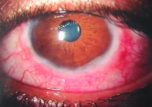
Corneal involvement is what turns this from an uncomfortable disease into a sight-threatening one. Superficial punctate keratopathy is common in active disease.
In more severe or poorly controlled cases, a shield ulcer, a sterile, oval epithelial defect typically in the superior cornea, can develop from mechanical trauma by overlying giant papillae combined with eosinophil-mediated epithelial toxicity, and this can scar and threaten vision if it is not recognised and treated promptly.
Pseudogerontoxon, a grey, arc-shaped limbal scar resembling arcus senilis but in a young patient, is a residual marker of previous limbal inflammation and, once present, does not itself require treatment.
Differential Diagnosis
- Seasonal or perennial allergic conjunctivitis, generally milder, without giant papillae or corneal involvement, and responsive to standard antihistamine and mast cell stabiliser therapy
- Giant papillary conjunctivitis, typically related to contact lens wear or an exposed suture, with a relevant history that VKC lacks
- Atopic keratoconjunctivitis, which tends to present in an older age group, often with associated eyelid skin changes and a more chronic course extending well beyond childhood
- Superior limbic keratoconjunctivitis, which involves the superior bulbar conjunctiva rather than the tarsal conjunctiva and lacks the giant papillae characteristic of VKC
Diagnostic Evaluation
Diagnosis is primarily clinical, based on the characteristic history and slit-lamp findings described above. Conjunctival scraping showing eosinophils can support the diagnosis when the picture is not entirely clear, though it is not required in a typical presentation.
Corneal examination with fluorescein staining is important in every case to look specifically for punctate keratopathy or shield ulceration, since the degree of corneal involvement, not the severity of itching, is what should drive treatment intensity.
Management
Cold compresses and preservative-free artificial tears provide symptomatic relief and are reasonable first steps in mild disease. Topical antihistamines and mast cell stabilisers, or combination agents, form the mainstay of treatment for mild to moderate disease and are generally well tolerated for long-term use, including outside the active season in perennial or high-risk patients.
Topical corticosteroids are necessary for moderate to severe exacerbations and are the most effective agents for controlling acute inflammation, but they carry real risks with prolonged use, including cataract and steroid-induced glaucoma, particularly relevant given how young these patients typically are and how many years of potential exposure lie ahead of them.
The general approach is to use the lowest effective dose for the shortest necessary course, with a clear plan to step down once the acute episode is controlled, rather than maintaining a child on long-term steroid drops by default.
Topical calcineurin inhibitors, particularly ciclosporin, have become an important steroid-sparing option for chronic or recurrent disease, allowing many patients to reduce their reliance on corticosteroids substantially. Tacrolimus ointment is used similarly in some centres.
A shield ulcer needs prompt, aggressive treatment, including topical steroids and sometimes a bandage contact lens or, in severe cases, surgical debridement, since delayed treatment risks permanent corneal scarring at the visual axis.
Prognosis
VKC tends to improve with age and frequently resolves or becomes substantially milder by late adolescence or early adulthood, which is genuinely reassuring information for families managing a young child through repeated flares.
The main determinant of long-term visual outcome is corneal involvement during the active disease years: patients who develop shield ulcers or significant keratopathy are at risk of permanent scarring and reduced vision if these complications are not caught and treated promptly, while patients whose disease stays limited to the conjunctiva generally do well long-term regardless of how uncomfortable individual flares have been.
Ongoing follow-up through the paediatric and adolescent years, with attention to corneal findings at every visit rather than symptom severity alone, is what keeps this disease from causing the vision loss it is capable of.


Document what you see
Two smartphone imaging tools built for everyday clinic use — one for the slit lamp, one for the fundus.
From Choroida — the team behind this siteReferences
- Bonini S, Coassin M, Aronni S, Lambiase A. Vernal keratoconjunctivitis. Eye. 2004.
- Leonardi A. Management of vernal keratoconjunctivitis. Ophthalmology and Therapy. 2013.
- Sacchetti M, Baiardini I, Lambiase A, et al. Vernal keratoconjunctivitis-related quality of life in children and adolescents. Pediatric Allergy and Immunology. 2018.
- Vernal Keratoconjunctivitis. EyeWiki, American Academy of Ophthalmology.
- Vernal Keratoconjunctivitis. StatPearls, NCBI Bookshelf.
Test yourself
A few questions straight from this article.
-
Which demographic pattern is typical of vernal keratoconjunctivitis?
VKC is a chronic bilateral allergic conjunctivitis with clear male predominance in childhood, usually beginning before the age of ten. The sex difference tends to narrow or disappear by adulthood. -
Why does vernal keratoconjunctivitis respond less reliably to antihistamines than simple allergic conjunctivitis?
VKC involves both type I IgE-mediated hypersensitivity and a chronic cell-mediated component. That second arm is why it behaves differently from a simple type I reaction and needs more than antihistamines. -
Which mediator is thought to be principally responsible for corneal epithelial damage in vernal keratoconjunctivitis?
Eosinophil proteins including major basic protein are directly toxic to the corneal epithelium. This is thought to drive the epithelial defects and shield ulcers, with mechanical rubbing from giant papillae contributing as well. -
Which finding characterises the palpebral form of vernal keratoconjunctivitis?
The palpebral form shows giant papillae with a cobblestone appearance on the superior tarsal conjunctiva. It is the form most associated with mechanical corneal complications, as the papillae rub the ocular surface with every blink. -
Horner-Trantas dots at the limbus represent aggregates of which material?
These small white points are aggregates of degenerated eosinophils and epithelial cells at the limbus. They accompany the gelatinous limbal thickening that defines the limbal form of VKC. -
How is a shield ulcer in vernal keratoconjunctivitis best described?
A shield ulcer is a sterile oval epithelial defect, typically superior, arising from mechanical trauma by overlying giant papillae plus eosinophil-mediated toxicity. It needs prompt aggressive treatment to avoid permanent scarring. -
Which features suggest atopic keratoconjunctivitis rather than vernal keratoconjunctivitis?
Atopic keratoconjunctivitis presents in an older age group, often with eyelid skin changes, and follows a more chronic course extending well beyond childhood. VKC by contrast tends to settle by early adulthood. -
What should drive the intensity of treatment in vernal keratoconjunctivitis?
Fluorescein examination for punctate keratopathy or shield ulceration matters in every case, because corneal involvement rather than symptom severity should set treatment intensity. Follow-up should check the cornea at every visit. -
Which agent has become an important steroid-sparing option for chronic vernal keratoconjunctivitis?
Topical calcineurin inhibitors, particularly ciclosporin, let many patients reduce corticosteroid reliance substantially; tacrolimus ointment is used similarly in some centres. This matters because prolonged steroids risk cataract and steroid-induced glaucoma in very young patients. -
What is the typical natural history of vernal keratoconjunctivitis?
VKC tends to improve with age and frequently resolves or becomes much milder by late adolescence or early adulthood. Long-term vision depends on whether corneal complications occurred during the active years.

Writes and reviews the clinical articles on Educate. Every post is checked against peer-reviewed sources before it goes live.


