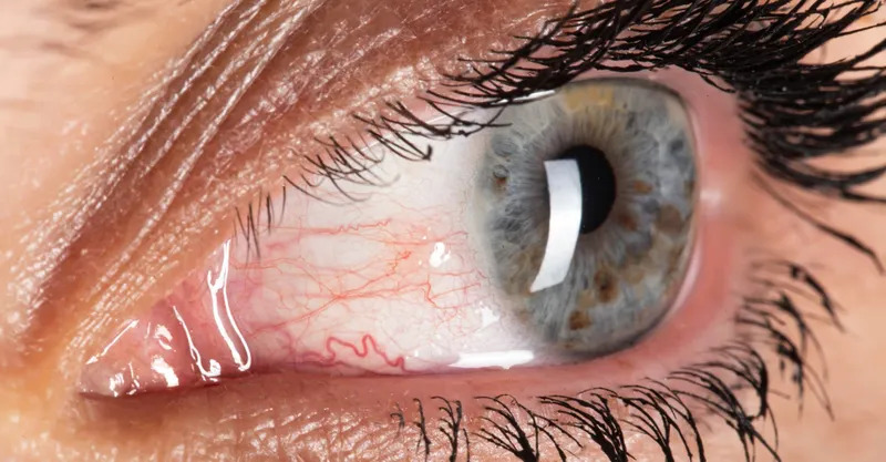
Episcleritis is a benign inflammation of the episclera, the thin vascular layer between the conjunctiva and the sclera.
It produces a sectoral or diffuse red eye with mild discomfort and normal vision.
Therefore, the key task is to confirm that the eye is not scleritis.
What Episcleritis Is
The episclera lies just above the sclera and carries a superficial and a deep vascular plexus.
In episcleritis, the superficial plexus becomes engorged and the tissue swells slightly.
Also, the inflammation stays on the surface and does not damage the scleral wall.
Because the disease is self-limited, most patients need little or no treatment.
Simple Episcleritis
The simple form is the most common and presents with sudden sectoral or diffuse redness.
Patients report burning, grittiness or a dull ache, but not true pain.
Also, the redness often peaks within hours and fades over one to two weeks.
Because the eye is otherwise quiet, vision and the pupil stay normal.
All-fit Slit-Lamp Adapter
Record and share exactly what you see at the slit lamp. One adapter fits any slit lamp or surgical microscope — and any smartphone.
From Choroida — the team behind this siteNodular Episcleritis
The nodular form shows a raised, mobile, tender nodule surrounded by injected vessels.
The nodule moves over the sclera when a cotton swab pushes it.
Also, it lasts longer than the simple form and may take several weeks to settle.
In contrast, a scleral nodule is fixed and lies deeper (see scleritis).
The Phenylephrine Test
First, examine the eye in daylight, since the shade of the redness matters.
Episcleritis looks bright red or salmon pink, while scleritis has a violet hue.
Then, instill one drop of 2.5% phenylephrine and look again after ten minutes.
The superficial vessels blanch in episcleritis, but the deeper scleral vessels do not.
Episcleritis Versus Scleritis
Pain is the best single clue, since scleritis causes a deep, boring pain that wakes the patient at night.
In addition, the globe is tender and the pain may spread to the brow or jaw.
Also, scleritis can thin the wall and reduce vision (see scleritis classification).
Therefore, when the pain is severe, treat the eye as scleritis until proven otherwise.
Differential Diagnosis of the Red Eye
Conjunctivitis has discharge, papillae or follicles and affects the whole surface.
Dry eye and blepharitis cause diffuse redness with lid or tear film signs (see dry eye disease and blepharitis).
Also, pingueculitis and phlyctenulosis produce a localized, raised lesion near the limbus.
In addition, herpes zoster can start as a red eye before the rash (see herpes zoster ophthalmicus).
Causes and Systemic Associations of Episcleritis
Most cases are idiopathic, and no cause is ever found.
However, a minority associate with rheumatoid arthritis, lupus, spondyloarthropathy, inflammatory bowel disease, gout or rosacea.
Also, herpes viruses and, rarely, syphilis or tuberculosis can be responsible.
Because the link is often mild, the systemic disease may already be known.
When to Investigate
A first, mild episode in a healthy adult needs no tests.
In contrast, recurrent, bilateral or nodular disease deserves a short workup.
Also, ask about joint pain, back pain, bowel symptoms, rash and dry mouth.
Then choose tests that match the history, such as a blood count, ESR, CRP, antinuclear antibodies, rheumatoid factor and syphilis serology.
Treatment of Episcleritis
Reassure the patient and advise cold compresses and preservative-free artificial tears.
Because the condition settles on its own, treatment aims at comfort.
Also, a short course of a topical NSAID or a low-potency steroid may speed recovery.
In nodular or painful cases, an oral NSAID such as flurbiprofen is often effective.
However, prolonged steroid use can raise the pressure and cause cataract, so avoid repeated courses.
Course and Recurrence of Episcleritis
A simple attack resolves in one to two weeks, and the nodular form in up to six.
Recurrences are common, often at intervals of weeks or months.
Also, stress, hormonal change and dry air may precede an attack.
Because episodes rarely scar, the long-term outlook for vision is excellent.
Follow-Up and Red Flags
Review the patient if there is no improvement after two weeks.
Return visits are also needed for rising pain, blurred vision or a bluish hue.
In addition, sudden loss of vision or a white thinned patch needs urgent assessment.
Therefore, give every patient clear written instructions.
An Example Case
A 32-year-old woman presents with a sectoral red patch in her right eye for two days.
She has mild burning, no discharge and a visual acuity of 20/20.
After 2.5% phenylephrine, the vessels blanch completely and the sclera is not tender.
The diagnosis is simple episcleritis, and cold compresses and tears are enough.
For a published case presentation, see the episcleritis case report.
Pitfalls to Avoid
- Do not call a painful, tender red eye episcleritis before checking the phenylephrine response
- Never prescribe long courses of topical steroids without reviewing the eye
- Do not skip the systemic history in recurrent or nodular disease
- Never miss herpes zoster ophthalmicus in a patient with a unilateral red eye and forehead pain
For the latest studies, see the PubMed results for episcleritis.


Document what you see
Two smartphone imaging tools built for everyday clinic use — one for the slit lamp, one for the fundus.
From Choroida — the team behind this siteReferences
- Watson PG, Hayreh SS. Scleritis and episcleritis. Br J Ophthalmol. 1976;60:163-191.
- Akpek EK, Uy HS, Christen W, Gurdal C, Foster CS. Severity of episcleritis and systemic disease association. Ophthalmology. 1999;106:729-731.
- Sainz de la Maza M, Molina N, Gonzalez-Gonzalez LA, Doctor PP, Tauber J, Foster CS. Clinical characteristics of a large cohort of patients with scleritis and episcleritis. Ophthalmology. 2012;119:43-50.
- Lyons CJ, Hakin KN, Watson PG. Topical flurbiprofen: an effective treatment for episcleritis? Eye (Lond). 1990;4:521-525.

Writes and reviews the clinical articles on Educate. Every post is checked against peer-reviewed sources before it goes live.