Endothelial cell loss after cataract surgery is an unavoidable part of the operation, but its size varies a great deal.
The corneal endothelium does not regenerate, so each lost cell is lost for good, and the cornea stays clear only as long as enough cells remain.
In healthy eyes the loss is well tolerated, and in vulnerable eyes it can lead to corneal failure.
Why Endothelial Cell Loss After Cataract Surgery Matters
The endothelium is a single layer of cells on the back of the cornea that pumps fluid out of the stroma and keeps the cornea clear.
Also, healthy adults have about 2,500 cells per square millimeter, and the number falls slowly with age, by about 0.5 percent a year (see the references).
The cornea usually stays clear if the density remains above about 500 cells per square millimeter, though the threshold varies.
Endothelial Cell Loss After Cataract Surgery: How Much Is Lost?
Studies using specular microscopy find an average loss of about 4 to 10 percent in the first months after uncomplicated phacoemulsification, with larger losses in difficult cases (see the references).
The loss may continue at a rate higher than normal for years after surgery.
A loss of more than 15 to 20 percent is regarded as high.
All-fit Slit-Lamp Adapter
Record and share exactly what you see at the slit lamp. One adapter fits any slit lamp or surgical microscope — and any smartphone.
From Choroida — the team behind this siteEndothelial Cell Loss After Cataract Surgery: Causes of Loss During Surgery
- Ultrasound energy and turbulence, particularly with dense nuclei
- Fragments of the nucleus hitting the endothelium
- Contact with instruments, the IOL, or the phaco tip
- Irrigation flow and chamber instability
- Chemical effects of drugs and solutions
- Inflammation, and a rise in pressure after surgery
Dense cataracts need more energy and result in more loss.
Risk Factors of Endothelial Cell Loss After Cataract Surgery
- Fuchs endothelial dystrophy and low preoperative cell counts (see Fuchs endothelial corneal dystrophy)
- Dense brunescent cataracts (see brunescent cataract)
- Shallow anterior chamber and small eyes
- Pseudoexfoliation, glaucoma, and uveitis (see pseudoexfoliation syndrome)
- Previous surgery or trauma
- Long surgery with complications, such as posterior capsule rupture
- Older age and diabetes
Protecting the Endothelium
- Preoperative assessment: specular microscopy in at-risk eyes (see specular microscopy)
- Dispersive and cohesive viscoelastic, used in a soft-shell technique to coat the endothelium
- Phaco techniques that reduce energy, such as chop, with pulsed or burst modes
- Low flow and appropriate vacuum, with stable chamber depth
- Pre-softening a dense nucleus, and careful positioning of the tip
- Use of an intracameral dye or an iris hook to improve visibility and avoid tissue contact
- Considering a different technique such as manual small-incision surgery in very dense cases
In Fuchs dystrophy, plan for the chance of corneal decompensation, and discuss combined or staged endothelial keratoplasty when the count is low.
Consequences
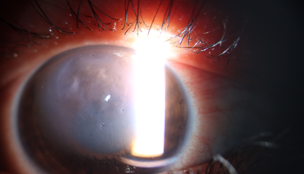
- Postoperative corneal edema, which may resolve in weeks
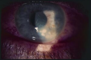
- Pseudophakic bullous keratopathy, a late complication, with permanent edema (see pseudophakic bullous keratopathy)
- Need for endothelial keratoplasty (see endothelial keratoplasty)
Counselling and Follow-Up
- Tell the patient before surgery if the endothelium is at risk, and explain the possibility of corneal swelling
- Examine the cornea at the first day, and review pachymetry or edema if present
- Measure cell counts at follow-up in high-risk eyes
- Manage pressure and inflammation early
Reading a Specular Microscopy Report
- Cell density: the number of cells per square millimeter
- Coefficient of variation: a measure of variability in cell size, with higher values meaning polymegethism
- Percent hexagonal cells: a measure of shape, which falls in stressed endothelium
- Central corneal thickness: a rise suggests that the pump is under strain
Compare the values before and after surgery, and use the same instrument.
An Example
A patient with early Fuchs dystrophy has a preoperative density of 1,400 cells per square millimeter and a dense cataract.
The surgeon uses a soft-shell technique, a low-energy chop method, and a stable chamber, and the density falls to 1,150 at three months.
The cornea clears by the second week, and the patient is followed with pachymetry.
Newer Approaches
Femtosecond laser fragmentation and low-energy techniques reduce the ultrasound energy used, and some studies show less endothelial loss in dense cataracts.
Evidence is still developing, and the choice depends on the equipment and the surgeon.
Long-Term Follow-Up
Cell loss continues at a higher rate for years in some eyes, so patients with a low count should be reviewed regularly for edema and for early signs of decompensation.
Use the same specular microscope, the same location, and the same method for each measurement, since small differences between devices can look like real loss.
Share the cell count with the patient when it is low, and explain that careful technique and follow-up can keep the cornea clear for many years in most cases.


Document what you see
Two smartphone imaging tools built for everyday clinic use — one for the slit lamp, one for the fundus.
From Choroida — the team behind this siteReferences
- Bourne WM, Nelson LR, Hodge DO. Central corneal endothelial cell changes over a ten-year period. Invest Ophthalmol Vis Sci. 1997;38:779-782.
- Bourne WM, Nelson LR, Hodge DO. Continued endothelial cell loss ten years after lens implantation. Ophthalmology. 1994;101:1014-1022.
- Walkow T, Anders N, Klebe S. Endothelial cell loss after phacoemulsification: relation to preoperative and intraoperative parameters. J Cataract Refract Surg. 2000;26:727-732.
- American Academy of Ophthalmology. Basic and Clinical Science Course, Section 11: Lens and Cataract. AAO; current edition.

Writes and reviews the clinical articles on Educate. Every post is checked against peer-reviewed sources before it goes live.