
Endothelial keratoplasty, encompassing both DSEK and DMEK, selectively replaces only the diseased endothelial layer of the cornea rather than the full-thickness graft required by traditional penetrating keratoplasty, discussed in its own dedicated article on this site.
This selective approach directly targets the layer actually responsible for most endothelial failure, sparing the patient’s own healthy stroma and epithelium and producing faster visual recovery with fewer refractive complications than a full-thickness transplant.
Understanding the difference between DSEK and DMEK, and why the field has moved increasingly toward the thinner DMEK technique, is central to understanding modern corneal transplant practice for endothelial disease.
DSEK: Descemet Stripping Endothelial Keratoplasty
DSEK transplants a thin disc of donor tissue consisting of posterior stroma, Descemet membrane, and endothelium, stripped from a donor cornea and inserted through a small incision into the recipient’s anterior chamber after the patient’s own diseased Descemet membrane and endothelium have been removed.
The donor tissue is unfolded and positioned against the posterior stroma, then held in place with an air or gas bubble while the endothelial pump reestablishes normal corneal deturgescence over the following days.
Because DSEK includes a thin layer of donor stroma along with the endothelium, the graft has more structural rigidity than a pure endothelial layer alone, making it technically somewhat easier to handle and position during surgery.
DMEK: Descemet Membrane Endothelial Keratoplasty
DMEK transplants only Descemet membrane and endothelium, without any donor stroma, making it a thinner, more anatomically precise replacement that most closely restores the eye’s original layered structure.
This thinness makes the tissue considerably more delicate and technically demanding to prepare, unfold, and position correctly in the recipient eye, and DMEK is generally considered to have a steeper learning curve than DSEK for the surgeon.
Despite the added technical difficulty, DMEK has become increasingly preferred where surgical expertise allows, given the visual outcomes discussed below.
Surgeons transitioning from DSEK to DMEK typically go through a deliberate learning curve, and many centers continue offering both techniques, matching the choice to the individual surgeon’s experience and the specific eye being treated.
All-fit Slit-Lamp Adapter
Record and share exactly what you see at the slit lamp. One adapter fits any slit lamp or surgical microscope — and any smartphone.
From Choroida — the team behind this siteWhy DMEK Is Increasingly Preferred
Because DMEK does not include any donor stroma, it avoids the stromal interface present in DSEK, an interface that can scatter light and modestly limit best-corrected visual outcomes even after a technically successful DSEK procedure.
Visual recovery after DMEK tends to be faster, and final visual acuity outcomes tend to be somewhat better on average compared to DSEK, reflecting this more precise anatomic replacement.
The rejection rate after DMEK is also lower than after DSEK, thought to relate to the smaller volume of donor tissue and the absence of donor stromal antigenic material, an advantage that matters considerably given how much endothelial keratoplasty is performed over a patient’s lifetime.
Indications
- Fuchs endothelial corneal dystrophy, discussed in its own dedicated article on this site, the most common indication for endothelial keratoplasty overall
- Pseudophakic bullous keratopathy, discussed in its own dedicated article on this site, from endothelial decompensation after cataract or other intraocular surgery
- Failed previous corneal graft with endothelial decompensation, where a repeat endothelial keratoplasty is often preferred over a repeat full-thickness graft
- Any other cause of isolated endothelial dysfunction with a healthy overlying stroma and epithelium, since the technique specifically depends on the rest of the cornea being structurally sound
Recovery and Complications
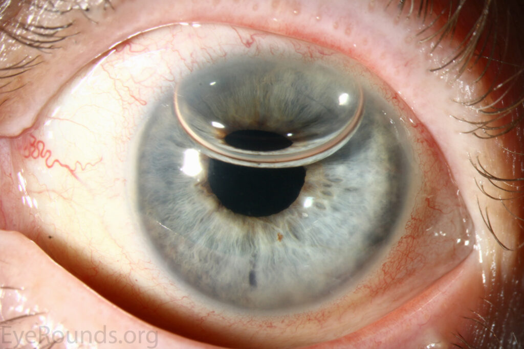
Graft detachment is the most common early complication specific to endothelial keratoplasty, since the thin donor tissue is held in place initially by an air or gas bubble rather than sutures, and a detached graft often requires a simple rebubbling procedure to reposition it.
Rejection, though less common than after penetrating keratoplasty and lower still with DMEK compared to DSEK, remains a lifelong risk requiring patient education about warning symptoms and prompt evaluation if they occur.
Visual recovery is generally faster than after penetrating keratoplasty, with many patients achieving useful vision within weeks rather than the many months sometimes required after a full-thickness graft, reflecting the absence of surface sutures and induced astigmatism.
Positioning the patient face-up for a period after surgery helps the air or gas bubble hold the graft in place against the posterior stroma, and clear instructions about this positioning requirement are an important part of preparing a patient before the procedure.


Document what you see
Two smartphone imaging tools built for everyday clinic use — one for the slit lamp, one for the fundus.
From Choroida — the team behind this siteReferences
- Price MO, Price FW Jr. Endothelial keratoplasty — a review. Clinical and Experimental Ophthalmology.
- American Academy of Ophthalmology. Basic and Clinical Science Course, Section 8: External Disease and Cornea.
- Dirisamer M, Ham L, Dapena I, et al. Efficacy of Descemet membrane endothelial keratoplasty: clinical outcome of 200 consecutive cases. Archives of Ophthalmology.
Test yourself
A few questions straight from this article.
-
What does endothelial keratoplasty replace in a cornea with endothelial failure?
Endothelial keratoplasty selectively replaces the layer responsible for the failure, sparing the patient's own healthy stroma and epithelium, unlike the full-thickness graft of penetrating keratoplasty. -
Which layers make up a DSEK graft?
DSEK transplants a thin disc of posterior stroma, Descemet membrane and endothelium, inserted through a small incision after the recipient's own Descemet membrane and endothelium are removed. -
Why is DSEK tissue easier to handle in surgery than DMEK tissue?
Because DSEK includes a thin layer of donor stroma with the endothelium, the graft has more structural rigidity than a pure endothelial layer, making it easier to handle and position. -
Why does DMEK tend to give better final visual acuity than DSEK?
DMEK contains no donor stroma, so it avoids the stromal interface present in DSEK, an interface that can scatter light and modestly limit best-corrected vision even after a technically successful DSEK. -
How does the rejection rate after DMEK compare with that after DSEK?
The rejection rate after DMEK is lower than after DSEK, thought to relate to the smaller volume of donor tissue and the absence of donor stromal antigenic material. -
What is the most common indication for endothelial keratoplasty overall?
Fuchs endothelial corneal dystrophy is the most common indication overall, followed by pseudophakic bullous keratopathy and failed previous grafts with endothelial decompensation. -
A previous corneal graft has failed from endothelial decompensation. What is often preferred?
Where a previous graft has failed with endothelial decompensation, a repeat endothelial keratoplasty is often preferred over a repeat full-thickness graft. -
What is the most common early complication specific to endothelial keratoplasty?
The thin donor tissue is held in place initially by an air or gas bubble rather than sutures, so graft detachment is the most common early complication and often needs a simple rebubbling to reposition it. -
Why is the patient positioned face-up after endothelial keratoplasty?
Face-up positioning lets the air or gas bubble hold the graft against the posterior stroma, and clear instructions about it are an important part of preparing the patient before surgery. -
How does visual recovery after endothelial keratoplasty compare with penetrating keratoplasty?
Many patients achieve useful vision within weeks rather than the many months sometimes needed after a full-thickness graft, reflecting the absence of surface sutures and induced astigmatism.

Writes and reviews the clinical articles on Educate. Every post is checked against peer-reviewed sources before it goes live.


