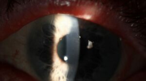
Hypopyon ulcer is a suppurative corneal infection with layered white cells in the anterior chamber.
The older term, ulcus serpens, describes a creeping ulcer classically linked to Streptococcus pneumoniae.
Because stromal destruction can progress within days, treat it as an emergency.
What Defines a Hypopyon Ulcer
A stromal infiltrate with an epithelial defect and layered exudate defines the condition.
However, hypopyon height does not identify the organism.
Also, review stromal corneal ulcers for the wider approach.
Ulcus Serpens: The Classic Picture
Classic ulcus serpens follows minor epithelial trauma, often with chronic dacryocystitis.
The ulcer then spreads with one advancing, undermined edge.
Stromal edema and Descemet folds often accompany a large hypopyon.
All-fit Slit-Lamp Adapter
Record and share exactly what you see at the slit lamp. One adapter fits any slit lamp or surgical microscope — and any smartphone.
From Choroida — the team behind this siteWhy the Hypopyon Forms
Toxins and inflammatory mediators diffuse through the stroma and inflame the iris and ciliary body.
Neutrophils then settle inferiorly in the anterior chamber.
In bacterial keratitis with intact Descemet membrane, the exudate is usually sterile.
However, fungi can penetrate Descemet membrane, so a fungal hypopyon may contain organisms.
See hypopyon for noninfectious causes.
Symptoms and Slit Lamp Exam in Hypopyon Ulcer
Patients report pain, photophobia, tearing and falling vision.
Therefore, ask about contact lens wear, trauma, vegetable matter and topical steroid use.
First, measure the infiltrate, epithelial defect and hypopyon height in millimeters.
Next, estimate stromal thinning and perform a Seidel test.
Finally, inspect the lacrimal sac, because chronic dacryocystitis can reseed the cornea.
Likely Organisms in Hypopyon Ulcer
S. pneumoniae is the classic cause of ulcus serpens.
Pseudomonas aeruginosa causes rapid necrosis with mucopurulent discharge, especially in lens wearers.
Meanwhile, Fusarium, Aspergillus and Candida lead the fungal differential.
Clues to Fungus or Acanthamoeba
Feathery margins, satellite lesions and an endothelial plaque suggest fungal keratitis.
Severe pain, a ring infiltrate and radial perineuritis point toward Acanthamoeba.
However, appearance alone cannot separate these organisms, so laboratory confirmation guides therapy.
Differential Diagnosis
For example, HLA-B27 uveitis and Behçet disease produce hypopyon without a suppurative infiltrate.
Peripheral lesions need separate consideration, including Mooren’s ulcer.
Finally, marked vitreous inflammation should prompt a B-scan to look for endophthalmitis.
Culture in Hypopyon Ulcer
Scrape before starting antimicrobials whenever this will not delay treatment.
Sample the leading edge and base, as described in corneal scraping.
Then send Gram and fungal stains with blood, chocolate and Sabouraud media.
Prior antibiotics reduce yield, so a negative culture does not exclude infection.
Empiric Treatment for Hypopyon Ulcer
Start intensive broad-spectrum topical therapy immediately after sampling.
Fortified cefazolin plus tobramycin is a common choice for large or deep ulcers.
Alternatively, a fourth-generation fluoroquinolone may serve where fortified drops are unavailable.
Add a cycloplegic for pain and to limit synechiae.
Because resistance varies by region, adjust therapy according to response and culture results.
Fungal Ulcer Treatment
When smear or history supports filamentary fungus, natamycin 5% is the usual first topical agent.
The Mycotic Ulcer Treatment Trial found better outcomes with natamycin than voriconazole, particularly in Fusarium ulcers.
However, deep disease responds slowly, so some patients need additional antifungal therapy or surgery.
Steroids in Hypopyon Ulcer
Avoid corticosteroids at presentation, while the organism remains unknown.
The SCUT trial tested topical prednisolone phosphate after 48 hours of moxifloxacin in bacterial ulcers.
It found no overall difference in three-month visual acuity.
However, subgroup analyzes suggested possible benefit in eyes with central ulcers or worse baseline vision.
Fungal ulcers fall outside this evidence.
Monitoring and Surgery
Review severe cases daily, and consider admission when adherence looks doubtful.
Useful signs include blunted margins, less density, epithelial healing and a shrinking hypopyon.
If the ulcer worsens, reassess the diagnosis and repeat cultures.
Progressive thinning calls for urgent consultation, because perforation may need tissue adhesive or therapeutic keratoplasty.
Counseling Patients With Hypopyon Ulcer
Explain that frequent drops continue overnight early on, because gaps allow progression.
Also, warn that central scarring can limit vision after the infection clears.
Tell patients to return at once for worsening pain or vision.
Pitfalls to Avoid
- Do not call the hypopyon sterile before excluding fungal infection
- Avoid starting steroids before identifying the organism
- Do not overlook the lacrimal sac in stubborn ulcers
- Never delay treatment while waiting for culture results
For the latest studies, see the PubMed results for hypopyon ulcer.


Document what you see
Two smartphone imaging tools built for everyday clinic use — one for the slit lamp, one for the fundus.
From Choroida — the team behind this siteReferences
- Lin A, Rhee MK, Akpek EK, et al. Bacterial keratitis Preferred Practice Pattern. Ophthalmology. 2019;126:P1-P55.
- Srinivasan M, Mascarenhas J, Rajaraman R, et al. Corticosteroids for bacterial keratitis: the Steroids for Corneal Ulcers Trial (SCUT). Arch Ophthalmol. 2012;130:143-150.
- Prajna NV, Krishnan T, Mascarenhas J, et al. The Mycotic Ulcer Treatment Trial: a randomized trial comparing natamycin vs voriconazole. JAMA Ophthalmol. 2013;131:422-429.
- Austin A, Lietman T, Rose-Nussbaumer J. Update on the management of infectious keratitis. Ophthalmology. 2017;124:1678-1689.
- McLeod SD, Kolahdouz-Isfahani A, Rostamian K, et al. The role of smears, cultures, and antibiotic sensitivity testing in the management of suspected infectious keratitis. Ophthalmology. 1996;103:23-28.

Writes and reviews the clinical articles on Educate. Every post is checked against peer-reviewed sources before it goes live.